Diagnosis of Arterial Thoracic Outlet Syndrome
Symptoms of Arterial TOS
Arterial TOS is rare and quite serious. The symptoms of arterial TOS are often sudden and severe. These symptoms are caused by sudden occlusion of one or more arteries in the affected arm. Arterial occlusion blocks blood flow to the arm, which may lead to severe complications if not diagnosed and treated promptly.

Symptoms of Arterial TOS
Patients with arterial TOS may present with acute arterial occlusion, local symptoms, intermittent arterial insufficiency, or chronic arterial insufficiency.
Patients with acute arterial occlusion may experience one or more of the 5 P’s:
- Pain
- Pallor
- Paresthesias
- Pulselessness
- Paralysis
Patients with these symptoms should seek immediate medical attention.
Local symptoms may include an aneurysm or a cervical rib. A patient may notice a cervical rib as a chronic hard lump at the base of their neck. A patient may feel a painless, pulsatile mass at the base of the neck. This pulsatile mass may represent a normal subclavian artery passing over a cervical rib, or a subclavian artery aneurysm.
Intermittent compression of the subclavian artery causes symptoms only in specific arm positions; in other words, symptoms only occur in those arm positions where extrinsic structures compress the artery. Some patients notice their arm “going to sleep” in certain arm positions, or when they wake up from sleep.
Chronic or intermittent compression of the artery may cause scar or stenosis of the artery. As a result, chronically limited blood flow could cause chronic arm fatigue. Under those circumstances of chronically limited blood flow, the patient would experience claudication.
Claudication is a symptom complex resulting from narrowing of a major artery, usually to an arm or a leg. Symptoms of claudication usually progress over time.
Clinical Diagnosis of Arterial TOS
Arterial TOS is rare, but may cause severe complications. The presentation of arterial TOS is dramatic and usually quite obvious. Doctors make the clinical diagnosis of arterial TOS promptly and accurately in most cases. Primary treatment of the arterial blood clot should begin almost immediately.

Physical Examination of Arterial TOS
Arterial TOS presents in a dramatic fashion, with potentially severe complications. Thus, a doctor would promptly begin with a physical examination focused on finding one or more of 4 pathologic processes.
Acute arterial occlusion puts the patient at risk of permanent tissue loss and loss of function. Because of this risk, a physician will always look for signs of acute arterial insufficiency, including the 5 Ps:
After this, the physician will look for signs of arterial emboli. These signs include skin ulcerations and discoloration, and splinter hemorrhages. Splinter hemorrhages are tiny blood clot fragments (emboli) that travel from the damaged artery to small arteries in the fingers. These emboli cause tiny hemorrhages where they lodge, which appear as tiny black spots. Thus, splinter hemorrhages are a sign of a proximal blood clot, usually in the arteries or heart. The presence of splinter hemorrhages necessitates a search for the source of emboli.
Your doctor will also examine the thoracic outlet on each side for the presence of a pulsatile mass. This mass may prove a subclavian artery aneurysm, or a normal subclavian artery passing over a cervical rib. With or without an aneurysm, your doctor may hear a bruit in the thoracic outlet with her stethoscope. A bruit is a swooshing sound caused by turbulent arterial blood flow. Narrowing of an artery may cause blood pressure in the two arms to be different. Finally, your physician will look for signs of chronic arterial insufficiency, including chronic skin changes.
Imaging Tests
Imaging tests are incredibly valuable tools in modern medicine. They are particularly valuable for a disease in a complex anatomic area like the thoracic outlet. In patients with arterial TOS, imaging tests confirm the doctor’s clinical diagnosis in several important ways. Read more about the value of imaging tests here.
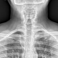 X-rays show excellent bone detail, but do not show soft tissue detail, and are limited to two dimensions. X-rays do not show arteries or veins. About 1% of the population has cervical ribs, but most of these people do not have arterial TOS. Even in patients with cervical ribs and arterial TOS, they have had cervical ribs their entire life, and only developed arterial TOS when they become symptomatic. X-rays thus have quite limited value in diagnosing arterial TOS and in planning treatment.
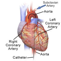 A flexible plastic catheter is inserted by a physician directly into an artery, usually in the groin. The physician advances the catheter under live x-ray (fluoroscopy) through the aorta, the main artery of the body, until it reaches the heart. At this point, the physician injects dye into the proximal aorta, after which the dye passes through the subclavian artery. As the dye passes through the arteries, x-rays create images. Direct angiography is an invasive test with a significant radiation dose. For this reason, CT angiography or MR angiography can almost always replace direct angiography.
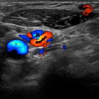 Ultrasound is performed without any radiation, and can demonstrate blood flow in the arteries in real time, allowing dynamic movement of the arms during the examination. However, bones create severe blind spots, limiting assessment of bony compression of arteries. Ultrasound is also quite limited for viewing fibrous bands and muscle anomalies.
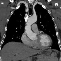 CT angiography is performed by injecting contrast into a peripheral vein, without an arterial catheter. CTA is fast, and shows excellent detail of bones and arteries. CTA requires significant radiation, and is limited in the evaluation of soft tissues, including fibrous bands and muscle anomalies. In cases of pure arterial TOS, CTA is the first-line test for many vascular surgeons.
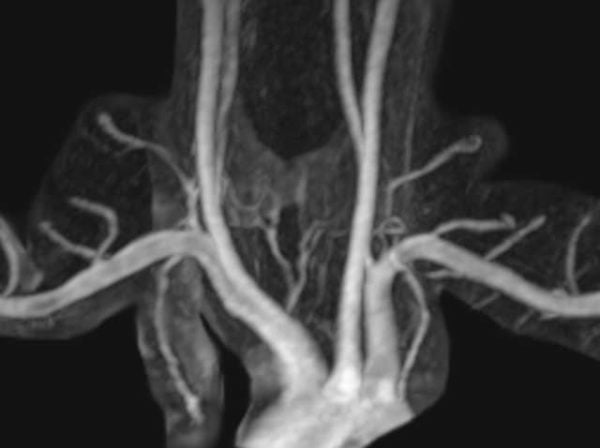
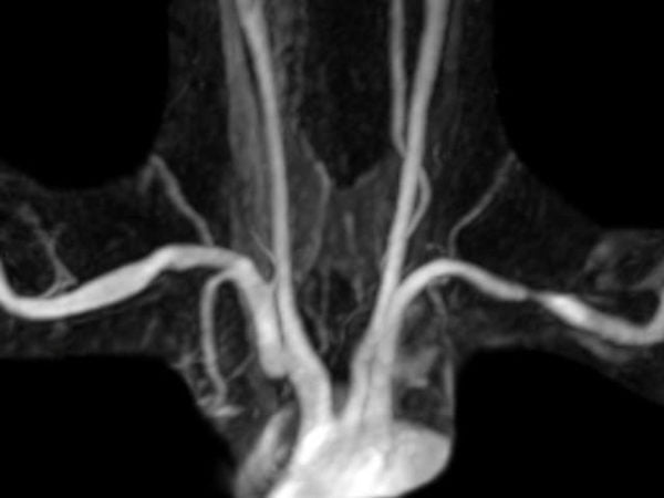
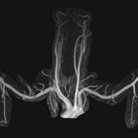 MR angiography is performed by injecting contrast into a peripheral vein, without an arterial catheter. MRA shows excellent arterial detail while eliminating most other structures, including bones. It requires no radiation. If used in conjunction with MRI, superb soft tissue detail is available. Excellent depiction of fibrous bands, muscle anomalies, bones, and the brachial plexus is possible with MRI.
Treatment
Critical treatment decisions revolve around the presence or absence of a blood clot causing arterial occlusion. When present, acute arterial occlusion demands urgent treatment. When absent, surgeons can assess and treat patients with arterial TOS less urgently.
Read more about the treatment of patients with arterial TOS here.
This interactive media demonstrates MR angiography images in two patients. Swipe between the two to demonstrate a normal MR angiogram and an MR angiogram with bilateral extrinsic compression.
Imaging Diagnosis of Arterial TOS
Venous TOS is quite uncommon. However, the presentation of venous TOS is quite dramatic, and complications can be serious. Venous TOS occurs when a blood clot forms in the main vein draining the arm. Although some treatment questions remain unsettled, doctors treat the blood clot urgently. Following this, doctors undertake diagnosis and treatment of the underlying cause.

Diagnosis
Your doctor will examine the thoracic outlet on each side for the presence of a pulsatile mass. This mass may prove a subclavian artery aneurysm, or a normal subclavian artery passing over a cervical rib. With or without an aneurysm, your doctor may hear a bruit in the thoracic outlet with her stethoscope. A bruit is a swooshing sound caused by turbulent arterial blood flow. Narrowing of an artery may cause blood pressure in the two arms to be different.
Because of the risk of permanent damage, your physician will always look for signs of acute arterial insufficiency, including the 5 Ps described above. Your physician will also look for signs of arterial emboli. These signs include skin ulcerations and discoloration, and splinter hemorrhages. Splinter hemorrhages are tiny emboli that travel from the damaged artery to small arteries in the fingers, causing tiny hemorrhages. Splinter hemorrhages are a sign of a proximal blood clot, usually in the arteries or heart. Their presence indicates the need for evaluation of the arteries or heart as a source of emboli. Finally, your physician will look for signs of chronic arterial insufficiency, including trophic skin changes, as described above.
Imaging Tests
Imaging tests are incredibly valuable tools in modern medicine. They are particularly valuable for a disease in a complex anatomic area like the thoracic outlet.
X-rays show excellent bone detail, but do not show soft tissue detail, and are limited to two dimensions. X-rays do not show arteries or veins. About 1% of the population has cervical ribs, but most of these people do not have arterial TOS. Even in patients with cervical ribs and arterial TOS, they have had cervical ribs their entire life, and only developed arterial TOS when they become symptomatic. X-rays thus have quite limited value in diagnosing arterial TOS and in planning treatment.
A flexible plastic catheter is inserted by a physician directly into an artery, usually in the groin. The physician advances the catheter under live x-ray (fluoroscopy) through the aorta, the main artery of the body, until it reaches the heart. At this point, the physician injects dye into the proximal aorta, after which the dye passes through the subclavian artery. As the dye passes through the arteries, x-rays create images. Direct angiography is an invasive test with a significant radiation dose. For this reason, CT angiography or MR angiography can almost always replace direct angiography.
Ultrasound is performed without any radiation, and can demonstrate blood flow in the arteries in real time, allowing dynamic movement of the arms during the examination. However, bones create severe blind spots, limiting assessment of bony compression of arteries. Ultrasound is also quite limited for viewing fibrous bands and muscle anomalies.
CT angiography is performed by injecting contrast into a peripheral vein, without an arterial catheter. CTA is fast, and shows excellent detail of bones and arteries. CTA requires significant radiation, and is limited in the evaluation of soft tissues, including fibrous bands and muscle anomalies. In cases of pure arterial TOS, CTA is the first-line test for many vascular surgeons.
MR angiography is performed by injecting contrast into a peripheral vein, without an arterial catheter. MRA shows excellent arterial detail while eliminating most other structures, including bones. It requires no radiation. If used in conjunction with MRI, superb soft tissue detail is available. Excellent depiction of fibrous bands, muscle anomalies, bones, and the brachial plexus is possible with MRI.
Interactive Media
This interactive media demonstrates magnetic resonance angiography (MRA) images in two different patients. One patient has a normal MRA. The other patient has compression of each subclavian artery. Note that the structures that cause this compression are not visible on MRA alone.