
History of Thoracic Outlet Syndrome
Table of Contents
Table of Contents
The Cervical Rib Syndrome
A cervical rib is an extra rib in the lower neck, above the normal first rib. Overall, cervical ribs are seen in about 0.5 to 1% of the human population. In detail, a cervical rib may be unilateral or bilateral, small or large, free-floating or fused to the first rib on the same side. While many physicians associate cervical ribs with thoracic outlet syndrome, the presence of a cervical rib does not prove or disprove thoracic outlet syndrome. Demonstration of cervical ribs goes back centuries, but association with TOS was only demonstrated in the early 19th century.

Galen
The cervical rib was first described in 150 A.D. by Galen. In general, dissection of the human body was forbidden at the time. However, Galen, the Greek anatomist and court physician to Marcus Aurelius, was allowed to do so, and published several texts on anatomy. In reality, Galen’s work was based in large part on dissections of apes. For several centuries, human dissection remained forbidden, and physicians learned anatomy only through Galen’s texts.
Andreas Vesalius
Andreas Vesalius is known as the father of modern anatomy. For a century and a half, Galen’s work had been accepted without question, and dissections of the human body had been essentially forbidden. Physicians learned human anatomy only by studying the texts of Galen. But Vesalius changed that.
Vesalius came from a family of prominent physicians, and immediately upon graduating from the University of Padua became the chair of Surgery and Anatomy. Vesalius replaced the reliance on Galen’s published textbooks by instituting the rigorous and meticulous direct study of the human body through dissection of cadavers. Often, Vesalius taught his students by performing dissections personally for them. Vesalius created large volumes of accurate drawings, and eventually published an updated version of Galen’s anatomic textbook, Institutiones Anatomicae.
Vesalius was criticized for this work, as Galen’s work was considered definitive and final. In fact, several years later, Vesalius established that Galen’s work had in fact been based on dissections of barbary apes rather than of humans. He then published a corrected version of Galen’s Opera omnia, which brought further criticism. Vesalius eventually published the seven-volume De humani corporis fabrica (On the fabric of the human body), now considered the foundation of modern anatomic study. Vesalius dedicated this work to Emperor Charles V, and eventually became Imperial physician of Charles’ court. Nonetheless, criticism of Vesalius’ work continued for many years.
Helkiah Crooke
Crooke described cervical ribs in man.
Helkiah Crooke served as Court physician to King James I of England. Crooke was also the first qualified doctor to be appointed Keeper of Royal Bethlem Hospital. This term Bedlam originated here.
Crooke had studied all the anatomy texts available in his day, and combined them with his personal experience and knowledge. Crooke had treated William Jaggard, who later published William Shakespeare. In 1615, Jaggard published Crooke’s 1000 page book, Mikrokosmographia: A Description of the Body of Man. Despite a significant amount of controversy, Crooke’s book became extremely popular, and was reprinted multiple times.
Crooke described cervical ribs thusly: “They are commonly both in men and in women on each side twelve, oftener more than fewer. For Nature would rather there should be an abundance than want. And in a publick, anatomy when a male factor was cut up, Bauhine found thirteen on each side; the first on the left side was perfect, but the first on the right side was imperfect. Fallopius also twice found one too many, Columbus once eleven at Padua.”
François Joseph Hunault
The cervical rib was again “rediscovered” by François Joseph Hunault in 1743. Hunault described and categorized supernumerary ribs in humans, including cervical ribs.
Sir Astley Cooper
Sir Astley Cooper was one of the leading surgeons of his day, publishing seminal work on the repair of inguinal hernias, and becoming renowned as a vascular surgeon. He was named sergeant surgeon to Kings George IV and William IV, and to Queen Victoria, and founded the famous medical school at Guy’s Hospital in London
Even though the cervical rib had been known for centuries, its effect on nearby structures was not documented until 1818, when Cooper described a young woman with pulselessness and gangrenous spots on her fingers, in conjunction with a palpable hard mass at the base of the neck. Cooper described, “I have however seen an exostosis arise from the sixth or seventh cervical vertebra, or perhaps from both” when introducing this case. Cooper attributed the patient’s symptoms to “a projection of the lower cervical vertebra towards the clavicle, and consequent pressure upon the subclavian artery.” Whether he in fact described a cervical rib or a C7 exostosis is unclear, but is a moot point. He had certainly presented the first description of arterial TOS.
Thus began the era of the Cervical Rib Syndrome.
H. Mayo
Mayo likely provided the first description of a subclavian artery aneurysm associated with a cervical rib.
As noted by Halsted in 1916, Mayo had described, “Right subclavian artery flattened. Unusual width.” Mayo’s description predated several other similar descriptions, including Adams in 1836, J. Mason Warren in 1849, and W.H. Willshire in 1860, who wrote, “…attention was drawn to a pulsation felt in the space corresponding to the subclavian triangle, first observed about six months ago, and which proves to be, so far as can be made out by examination, a supernumerary rib arising above the first…”.
John Hilton
John Hilton, a British surgeon, describes gangrene of the arm in a patient with compression of the subclavian artery caused by an exostosis of the first rib.
In 1849, Dr. Hilton became a full surgeon at Guy’s Hospital in London, and Professor of Anatomy and Surgery in the Royal College of Surgeons in 1860. Dr. Hilton performed one of the first recorded surgeries for an internal hernia with bowel strangulation, and was the first to use internal sphincterotomy for the treatment of anal fissure. The ‘White Line of Hilton’ he described is still a widely-recognized anatomic landmark for colorectal surgeons.
Holmes Coote
Holmes Coote performed the first TOS surgery.
A 26 year-old female servant described a hard mass in her left lower neck, present since childhood. The “tumor” had grown recently, and she experienced paresthesias in the tips of her fingers, with loss of pulses at the wrist, wasting of muscles in the arm, and weakness leading to dropping of items.
Mr. Coote explored the area while the patient was “fully under the influence of chloroform.” He noted a bony ‘tumor,’ which he had to divide from the cervical vertebra. He also found it was attached to the first rib anteriorly. He removed as much of the ‘tumor’ as he could, and pulses returned in the left upper extremity.
Coote described his trepidation at exploring this area thusly: “But the region was not a pleasant one for any proceeding demanding the use of the knife. The subclavian artery and vein were in front; the axillary plexus of nerves lay spread out above; below, the apex of the lung, covered by the pleura, rose up in dangerous proximity; on the scalenus was the phrenic nerve; while towards the mesial line were the important vessels and nerves passing to the head, together with the vertebral vessels and thoracic duct. You can understand, therefore, why I was cautious in what I did.”
The bony tumor was referred to as “an exostotic growth from the transverse process of the seventh cervical vertebra,” and “a development of the costal element, the rib, of the seventh cervical vertebra.”
Thus, Coote became the first person to surgically treat a TOS patient.
Gruber
Gruber published a classification system for cervical ribs. His classification was based on the length of the cervical rib and its attachment to the first rib.
Sir James Paget
In London in 1875, Sir James Paget first describes venous TOS.
Sir Paget is widely regarded as one of the two founders of modern pathology, along with Rudolf Virchow in Germany. He first established himself as a famous surgeon in London by mastering the major English, French, German, Dutch, and Italian medical literature of his time, and by applying a strict scientific discipline to the study of human physiology as it applied to surgery. Following this, in the field of pathology, Sir Paget’s work was groundbreaking. Sir Paget established the scientifically rigorous use of the microscope in the study of human infection and tumors. Following Paget, the microscope became the essential and indispensable scientific tool of pathology. Paget published Lectures on Surgical Pathology, one of the two landmark works in pathology. He served as surgeon extraordinary to Queen Victoria and surgeon in ordinary to the Prince of Wales, and was elected president of the Royal College of Surgeons.
Leopold von Schroetter contemporaneously and independently studied and described venous TOS, but did not publish until 1884.
Leopold von Schroetter
In Vienna, Leopold von Schroetter independently describes spontaneous thrombosis of the subclavian-axillary vein in young individuals.
Von Schroetter was an internist and laryngologist in Vienna. He developed the first modern lecture hall, with an accompanying laboratory. At the age of 78, he attended the Crown Prince of Germany.
H. Lewis Jones
In 1893, H. Lewis Jones described six patients with symmetric atrophy of hand muscles, usually preceded by pain and tingling. In 1906, he revisited these cases, along with additional cases he had seen since the original publication, and retrieved their x-rays. He found a strong correlation between symptoms and the presence of cervical ribs. In particular, he retrieved the records of 14 patients with pain and hand muscle atrophy. Of these 14, 10 had cervical ribs. 8 patients had bilateral symptoms and bilateral cervical ribs, and 2 had a unilateral cervical rib on the side of their symptoms.
In 1902, Farquhar Buzzard described 7 patients with brachial plexus involvement, at least one of which appears to have neurogenic TOS.
In 1903, Edwin F. Bramwell described an 18 year-old plumber with sensory and motor disturbances in his left arm. He found hand weakness and atrophy of intrinsic hand muscles. Bramwell described the abnormality as a “lesion of the first dorsal root.” Bramwell proposed the ‘sharp internal border of the first rib’ had caused nerve trauma.
Neither Buzzard nor Bramwell excluded cervical ribs in their patients.
While Jones did recognize the pattern of neurogenic TOS in young women as early as 1893, he did not confirm the association with cervical ribs until 1906. In the meantime, Thorburn had recognized the same association, and had already treated neurogenic TOS patients through the removal of cervical ribs by 1904.
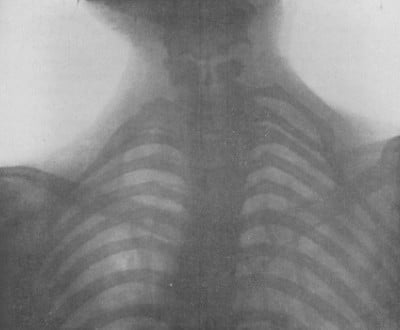
Wilhelm Conrad Roentgen
In 1895, Wilhelm Conrad Röntgen was working with cathode ray tubes when he discovered a novel type of electromagnetic radiation, which he called an “x-ray.” He noted that his body cast a strange image when he passed between the cathode ray tube and a barium-coated screen. After two weeks of experimentation, Röntgen created the first radiograph or roentgenogram. This famous image shows his wife’s hand with a ring.
The x-ray enabled doctors (and others) to see inside the living human for the first time, and became an international sensation. X-rays were used for many purposes, and in the setting of TOS made the diagnosis of cervical ribs easy and accurate. These images were known as ‘skiagrams,’ and became a standard part of the workup of patients presenting with vascular compression or aneurysm in the thoracic outlet.
John Benjamin Murphy
John Benjamin Murphy of Chicago is the first surgeon to resect a cervical rib associated with a subclavian artery aneurysm.
Dr. Murphy is still known for numerous surgical innovations, including Murphy’s button (to join two segments of bowel), Murphy’s sign (for the diagnosis of cholecystitis), Murphy’s tympani, and the Murphy test (for diagnosis of kidney disease). Dr. Murphy pioneered end-to-end anastomoses of hollow viscera and blood vessels, championed early appendectomy in cases of appendicitis (despite strong opposition from the Chicago medical establishment), and introduced therapeutic pneumothorax for pulmonary tuberculosis.
William Williams Keen
Dr. William Keen at Thomas Jefferson University in Philadelphia publishes a review of all 42 known cases of resected cervical ribs, and describes a clinical definition and surgical treatment for this disorder.
Dr. William Williams Keen became renowned as the first surgeon in America to remove a brain tumor and have the patient survive for 30 years. Dr. Keen had served as a surgeon in the Civil War, and utilized his experiences to publish the seminal book in its field, Gunshot Wounds and Other Injuries of the Nerves.
After the Civil War, Dr. Keen traveled to Europe to study with famed physicians Duchenne in Paris and Virchow in Berlin. Dr. Keen returned to Thomas Jefferson University in Philadelphia as professor of surgery, where he performed the first successful brain tumor removal, performed the first ventricular puncture for the treatment of hydrocephalus and increased intracranial pressure, and developed the technique of cervical nerve root section for the treatment of spasmodic torticollis. In 1887, Dr. Keen revised and re-edited the American edition of Grey’s anatomy, and in 1906 he became editor of the multivolume Surgery, Its Principles and Practice. In this volume, Dr. Harvey Cushing, with whom Dr. Keen had a long professional relationship, first published work that created a sensation in the medical world, and established the discipline of neurosurgery as a distinct field in medicine.
Physician to Presidents
In 1893, Dr. Keen assisted in removing a sarcoma from the mouth of President Grover Cleveland, an operation which was kept secret for more than 15 years, until after the President’s death. In 1921, Dr. Keen attended Franklin Delano Roosevelt after he contracted polio. Dr. Keen was the president of the American Medical Association in 1900, was the first surgeon to receive an honorary fellowship in the American College of Surgeons, was elected an honorary fellow of the Royal College of Surgeons in England and the Legion of Honor in France, and received honorary degrees from seven North American universities and four international universities.
J. B. Roberts
J. B. Roberts describes a series of different forms of cervical ribs, and proposes a surgical approach to each of these different forms.
Alfred Washington Adson and Jay R. Coffey
Adson and Coffey at the Mayo Clinic suggest the mechanism by which the anterior scalene muscle causes neurovascular compression in patients with cervical ribs, and are the first to demonstrate that scalenotomy alone can relieve the symptoms of neurovascular compression, without resection of the cervical rib. This procedure becomes popular for several decades, although the recurrence rate is very high.
Dr. Alfred Washington Adson created and headed the Section of Neurological Surgery at the Mayo Clinic. He was a pioneer in American surgery, was a founding member and president of the Society of Neurological Surgeons, and was president of the Minnesota State Medical Association and the Minnesota State Board of Medical Examiners..
Dr. Alfred Washington Adson created and headed the Section of Neurological Surgery at the Mayo Clinic. He was a pioneer in American surgery, was a founding member of the Society of Neurological Surgeons, and was president of the Society of Neurological Surgeons, of the Minnesota State Medical Association, and the Minnesota State Board of Medical Examiners.
In 1938, Adson performed the first total anterior scalenectomy, removing the entire muscle.
Cervical Rib Syndromes without a Cervical Rib
After the advent of the medical x-ray, physicians thought they had solved the problem of the cervical rib syndrome. Patients with vascular or neurologic symptoms underwent an x-ray examination, and a cervical rib appeared. Shortly after this issue had resolved, a number of reports arose in the medical literature regarding patients with cervical rib syndrome, in whom x-rays demonstrated no cervical rib. Now these same physicians found they faced an entirely new challenge-‘cervical rib syndrome without a cervical rib’ syndrome.
Edwin Bramwell and E. Farquhar Buzzard
Bramwell describes the first case of neurogenic TOS without a cervical rib.
An 18 year-old plumber developed sensory and motor disturbances in his left arm. Bramwell found weakness, with atrophy of intrinsic hand muscles. He described the abnormality as a “lesion of the first dorsal root.” Bramwell also proposed the ‘sharp internal border of the first rib’ had caused nerve trauma.
Bramwell also referred to several similar cases seen by Buzzard in 1902. Most of these cases appear to represent post-infectious neuropathy, although unusually limited to a single root. A final case noted in brief by Buzzard may, in fact, represent neurogenic TOS.
William Thorburn
First surgical removal of a cervical rib.
Thorburn describes two young female patients with cervical ribs on X-ray, and corresponding unilateral symptoms of diminished pulse, pain, hand muscle atrophy and weakness, and temperature sensory abnormalities. He removed the cervical rib in each of these patients, with considerable reduction of symptoms.
Thomas Murphy
Mr. Thomas Murphy, of Melbourne, Australia, performs the first resection of a normal first rib for the treatment of neurogenic thoracic outlet syndrome.
Thomas Murphy described a 28 year-old woman with symptoms of brachial plexus compression. X-rays showed no cervical rib, but Murphy was convinced the brachial plexus was compressed. Murphy performed surgery, finding the broad insertion of the anterior scalene was compressing the brachial plexus. He removed the insertion and a small part of the first rib. The patient had a “perfect and permanent” recovery.
Thomas Wingate Todd
Thomas Wingate Todd at Manchester University describes the potential causes of neurovascular compression in the thoracic outlet in a series of scientific papers outlining the anatomy and anatomical variations of the thoracic outlet. Dr. Todd’s anatomic dissections included descriptions of cervical ribs, anatomic variations of the first rib and scalene muscles, and the position of the clavicle with shoulder movements. Dr. Todd also postulated that the gradual descent of the shoulder girdle with aging causes narrowing of space between the clavicle and rib, contributing to neurovascular compression.
However, Todd also describes in detail a distinct mechanism causing narrowing of only small, distal arteries. In 1912, he shows sympathetic nerve fibers passing from the T2 nerve root to the T1 nerve root variably in humans. In 1913, he demonstrates that pressure on sympathetic nerves causes narrowing and occlusion of small arteries.
Dr. Todd became Professor and Chairman of Anatomy at what is now Case Western Reserve University Medical School in Cleveland in 1912. Dr. Todd built the Hamann-Todd Osteological Collection, the world’s largest collection of human and anthropoid skeletons. He also publishes The Atlas of Skeletal Maturation in 1937, and initiated the Brush study in 1926, both of which are widely-accepted tools for the evaluation of human skeletal growth and maturation.
William Halsted
William Halsted publishes an article reviewing the available knowledge regarding subclavian artery aneurysms associated with cervical ribs. He then tried to recreate and explain the mechanism.
Halsted collated 716 cases of cervical ribs from the medical literature, and found 27 with subclavian artery aneurysms. He performed experiments on dogs, creating arterial constrictions of varying degrees for varying lengths of time until he reproduced an aneurysm appearing immediately distal to the constriction. Halsted then proposed a mechanism by which such aneurysm formation could occur.
Dr. Halsted was one of the pioneers of modern surgical technique in the United States, using aseptic technique and novel wound closure techniques to advance the safety and efficacy of surgery. He performs the first emergency blood transfusion, the first radical mastectomy, the first nerve block, the first inguinal hernia repair, and invents the surgical glove. Dr. Halsted was named the first surgeon-in-chief of the Johns Hopkins Hospital, and was the first professor of surgery at Johns Hopkins Medical School. He created subspecialty divisions in the surgery department, and the radiology department at Johns Hopkins. Dr. Halsted became renowned for training surgical residents, many of whom went on to create surgical residency programs at institutions throughout the United States, spreading the prodigious influence of Dr. Halsted. Dr. Halsted was also the captain of the first American 11-player football team in 1870.
John Sebastian Bach Stopford and E. D. Telford
John Sebastian Bach Stopford and E. D. Telford at Manchester University Medical School describe compression of the lower trunk of the brachial plexus by a normal first rib, and report results from 10 patients following resection of the first rib.
Stopford and Telford considered the following three key elements as the underlying etiologic factors in development of brachial plexus compression:
Stopford and Telford reported the first case of a patient with neurogenic symptoms and a cervical rib, in whom successful treatment was performed by resection of the normal first rib, leaving the cervical rib in place.
John Sebastian Bach Stopford trained under Thomas Wingate Todd, and wrote numerous scientific papers on the traumatic disruption of nerves, the regeneration of injured nerves, the autonomic nervous system, and the compression of peripheral nerves, most notably that caused by cervical ribs. Dr. Stopford’s monograph on the circulation of the pons and medulla is regarded as a classic publication. Later in his career, Dr. Stopford served as the Chair of Anatomy, the Dean of the medical school, and the Vice-Chancellor of the University. Dr. Stopford was appointed M.B.E. and K.B.E., was knighted in 1941, was awarded honorary degrees in science and law at the University of Cambridge and other universities, and was awarded Honorary Fellowships in the Royal College of Surgeons and the Royal College of Physicians.
Stopford and Telford made further contributions to our knowledge of TOS. Please see their additional timeline entry for 1931.
Arthur Ayer Law
Dr. Arthur Ayer Law demonstrates the existence of several soft tissue bands that cause neurovascular compression, simulating a cervical rib.
Gould, Symonds, Yates and Guest
First cases of stroke caused by arterial TOS.
Gould described a 19 year-old laborer who had pain, weakness and coldness of his right hand. Gould found a cervical rib on examination, and he could create turbulence within the adjacent subclavian artery by pressing on it. Years later, the right arm pulse disappeared and weakness appeared in the left arm (likely due to stroke).
In 1927, C.P. Symonds described two patients presenting with left-sided weakness, who also had right-sided cervical ribs and arterial TOS. Dr. Symonds proposed a mechanism tying the two processes together. Symonds suggested that the cervical rib caused damage to the right subclavian artery, resulting in blood clot formation. This blood clot extended proximally towards the heart until it entered the right common carotid artery. At this point, clot fragments broke off and traveled to the brain, causing stroke.
Neither author provided pathologic proof in these patients, although Symonds confirmed cervical ribs with x-rays.
In 1928, Yates and Guest described a patient with a non-united right clavicle fracture, loss of pulses and weakness. The patient suffered a stroke which resulted in their death. Autopsy showed the following:
This case provided pathologic proof of the process proposed by Symonds.
Howard Christian Naffziger and Francis Clark Grant
Naffziger and Grant first introduce the concept of neurovascular compression in the thoracic outlet due to scalene muscle anomalies, without the presence of a cervical rib. They perform the first scalenotomies in patients without cervical ribs, but do not publish their findings until 1937 and 1938.
Dr. Howard Christian Naffziger, who trained under eminent surgeons William Halsted and Harvey Cushing, became one of the most esteemed neurosurgeons of his time. He created the division of Neurosurgery and served as Chairman of the Department of Surgery at the University of California San Francisco, was elected president of the American College of Surgeons, and was Chairman of the committee that established the American Board of Neurological Surgeons.
Francis Clark Grant trained under eminent neurosurgeons Charles Frazier and Harvey Cushing. Dr. Grant succeeded Dr. Frazier as Professor and Chairman of Neurosurgery at the School of Medicine and the University Hospital at University of Pennsylvania. He published over 200 papers in his lifetime, and refined or developed several neurosurgical procedures and instruments.

John Sebastian Bach Stopford and E. D. Telford
Stopford and Telford describe a new mechanism of arterial occlusion caused by nerve pressure in the thoracic outlet, without involvement of the subclavian artery. They describe three patients with severe arterial occlusion and distal gangrene of the fingers and hand. Each of these patients had a cervical rib, but no subclavian artery compression or aneurysm. And, in each patient, removal of the cervical rib provides complete relief of symptoms.
Specifically, Stopford and Telford explain how sympathetic fibers of the lower brachial plexus regulate the small arteries of the arm and hand. Considering prior work by Wingate, they demonstrate how some patients have variable anatomy of these fibers, immediately adjacent to the first rib. Thus, pressure by the first rib, or a cervical rib, in some patients causes abnormal regulation of the small arteries. As a result, these arteries constrict over a long period of time. At first, the patients experience pallor and coldness from loss of arterial blood flow. Over time, the lining of the arteries expands, blocking arterial flow. This results in gangrene, or loss of tissue.
Alton Ochsner, Mims Gage and Michael DeBakey
Renowned surgeons Alton Ochsner, Mims Gage and Michael DeBakey at LSU publish a comprehensive study of patients with symptoms of neurovascular compression in the thoracic outlet in the absence of a cervical rib, for which they coin the term, “Scalenus Anticus Syndrome”. They credit Naffziger with first recognizing this mechanism in 1929 in patients without cervical ribs, and suggest the term “Naffziger Syndrome”.
Dr. Alton Ochsner was named Chairman of Surgery at Tulane Medical School at the young age of 31, and founded the world-famous Ochsner clinic at Charity Hospital in New Orleans, which remains one of the pre-eminent surgical teaching programs in the country. Dr. Ochsner was the first to report the link between cigarette smoking and lung cancer, and he trained some of the most prominent surgeons of the time, including Dr. Michael DeBakey.
Dr. Michael DeBakey is one of the most renowned cardiovascular surgeons in the world. After volunteering for military service in World War II, Dr. DeBakey created the concept of the Mobile Army Surgical Hospital (M*A*S*H unit) that enjoyed stellar success during the Korean War. Dr. DeBakey was one of the first cardiothoracic surgeons to perform coronary bypass surgery, was the first man to perform carotid endarterectomy, and made numerous other contributions to and innovations in cardiovascular surgery, including work on the Dacron artificial graft, the heart-lung machine, and the artificial heart.

Alfred Washington Adson
Adson performs the first scalenectomy, or resection of the entire anterior scalene muscle. This procedure is then performed intermittently over the next several decades, until Sanders introduces a more refined technique in 1979.

Kenneth C. Eden
In 1939, Eden first demonstrated the role of the clavicle in TOS.
Eden operated on three patients with aneurysm or occlusion of the subclavian artery; two with a cervical rib, and one without. At surgery, he demonstrated marked narrowing of the space below the clavicle on arm motion. In patients with a cervical rib or a normal first rib, arterial damage occurred in this narrowed space.
Eden stated, “The findings in these three cases suggest that the wall of the subclavian artery is weakened by intermittent compression by the clavicle against a bony obstruction. This is usually provided by…a complete cervical rib, but an abnormal first thoracic rib may do so also.”

Murray Falconer and Graham Weddell
Murray Falconer and Graham Weddell describe military recruits with neurovascular compression. These recruits experience symptoms while carrying heavy backpacks or assuming the “military” position, with the shoulders thrust backwards. Falconer and Weddell propose that the weight depresses the shoulders and causes neurovascular compression between the normal clavicle and first rib. Falconer and Weddell coin the term “costoclavicular syndrome”. Note that Eden first considered this mechanism in 1939.
Controversy about the existence of costoclavicular syndrome developed. In 1962, Falconer and Dr. Franklin W. P. Li described a series of 11 patients who underwent first rib resection with good results. Falconer and Li felt that the success of their approach confirmed the mechanism of costoclavicular syndrome. Several of these patients had been previously unsuccessfully treated for carpal tunnel syndrome.
Dr. Falconer was one of the strongest proponents for the use of surgical ablation in the treatment of temporal lobe epilepsy. He performed a fellowship under pioneer neurosurgeon Alfred Adson at the Mayo clinic in 1937, and was named first Director of Neurosurgery at Guy’s Hospital in London in 1949. Dr. Falconer developed the renowned program at this institution for the treatment of temporal lobe epilepsy, and over his career helped to elucidate the pathology of temporal lobe epilepsy, and developed and refined the surgical criteria and techniques for treatment of patients with this condition.
R. L. Swank
R. L. Swank first describes the differences between upper and lower brachial plexus involvement in thoracic outlet syndrome.

Irving S. Wright
Irving S. Wright describes patients with similar symptoms and signs as those with ‘costoclavicular syndrome.’ However, Wright’s patients experience symptoms with hyperabduction of the arms (elevation of the arms above the head). Interestingly, Wright could not reproduce symptoms or pulse obliteration in his patients by using the maneuvers used by Falconer.
Wright went on to test 150 asymptomatic volunteers by performing hyperabduction of their arms while monitoring their pulse. In nearly 90% of volunteers, Wright induced obliteration of the radial pulse, despite the volunteers having no symptoms. In only two of the 150 patients could Wright induce tingling, presumed to represent nerve compression. Interestingly, neither of these patients had a loss of pulse in the same arm position. Wright concluded that clinical obliteration of the pulse was not correlated with compression of the brachial plexus.
Wright also performed anatomic dissections to further understand the underlying mechanism of hyperabduction. He proposed that there were two different mechanisms, which might occur in isolation or in combination in different patients. First, Wright proposed that the space posterior to the pectoralis minor muscle would create “stretching, torsion, and pinching” of the vessels and brachial plexus only in hyperabduction. Second, he proposed that different arm motions, as demonstrated by Eden in 1939 and Falconer in 1943, would cause similar changes between the clavicle and the first rib.
Finally, Wright suggests that these mechanical factors cause two different types of pathologic process: “The stretching or pinching of the nerve trunks probably produces the immediate paresthesias. Ischemia due to impaired blood supply to the nerves may play a part after prolonged hyperabduction.”
A Recognized Pioneer
Dr. Wright is known as a pioneer in the treatment of blood clots. Blood clots in the legs can break off and lodge in the lungs. These pulmonary emboli may be fatal. Heart attacks are caused by blood clots in the coronary arteries. Dr. Wright had suffered months of severe blood clots in his 30s. After recovering, he used an experimental drug called Heparin to successfully treat a patient with similar disease. Dr. Wright was awarded the prestigious Albert Lasker Award from the American Heart Association in 1960, recognizing his study of Heparin use in 800 patients with heart attacks. Dr. Wright served as president of the American Heart Association, the American College of Physicians and the American Geriatric Society.
Jere Lord
Dr. Jere W. Lord performs the first claviculectomy (removal of the clavicle). Lord aims to relieve compression of the neurovascular bundle in the costoclavicular interval. Unfortunately, many authorities consider this procedure cosmetically disfiguring, and feel it causes a significant alteration in function, and it never becomes popular.
Dr. Lord was a professor of clinical surgery at New York University School of Medicine, a present of the New York Heart Association, and he published over 180 papers in the academic literature.
Dr. Lord referred to Telford and Mottershead in 1948, and stated, “If one analyzes the anatomical structures through which the subclavian and axillary artery and vein must pass, then it is evident that no single operative procedure can be successful in all cases of the shoulder girdle syndromes.” This prescient statement remains pertinent today.

Jere Lord and Peter Stone
Lord and Stone publish the first report on pectoralis minor tenotomy.
They perform pectoralis minor tenotomy in addition to anterior scalenectomy in 5 patients, with good results in 4 patients.
Unification of Thoracic Outlet Syndromes and the Modern TOS Era
As researchers and anatomists learned about the numerous anatomic variations and causes of thoracic outlet syndrome, it became apparent that many patients suffered from related but different clinical syndromes. A number of authors published articles regarding these syndromes, along with a number of different provocative maneuvers. In 1956, Peet created an umbrella term to encompass all of these variable syndromes, and ‘thoracic outlet syndrome’ was born.

R. M. Peet
R. M. Peet and colleagues first use the term “thoracic-outlet syndrome” to unify the large number of upper extremity neurovascular compression syndromes.
In 1958, Rob and Standeven describe a series of patients with cervical ribs, arterial thromboses or emboli, and distal gangrene of the upper extremity. They use the term, “thoracic outlet compression syndrome” for the first time.
O. Theron Claggett
O. Theron Claggett, the president of the American Association for Thoracic Surgery, and one of the premiere thoracic surgeons of the mid-twentieth century, re-introduces the first rib resection after decades of disappointing results from scalenotomy alone. In his presidential speech to the American Association for Thoracic Surgery, Claggett recounts an extensive history of TOS, and proposes first rib resection by the posterior approach. This posterior approach eventually falls out of favor because it requires a fairly radical soft-tissue dissection, and because it creates disruption of the posterior shoulder girdle musculature, which is now felt to be important in the stability of the shoulder girdle.

1960s
A number of authors publish descriptions of supraclavicular and infraclavicular approaches for first rib resection, but these approaches are not widely accepted.
David Roos
Dr. David Roos performs the first transaxillary first rib resection. Dr. Roos demonstrates and categorizes numerous soft tissue anomalies in the thoracic outlet. His transaxillary approach goes on to achieve wide acceptance, due to a less radical soft-tissue dissection than previous approaches, and to improved visualization of the muscular and fibrous anomalies that are known to exist in the thoracic outlet.
D. Silver and Chandu Vemuri
D. Silver recommends tenotomy of the pectoralis minor in selected cases of first rib resection. Vemuri et al suggest that a population of neurogenic TOS patients have predominant compression of the brachial plexus in the retropectoralis space. Thus, these authors suggest pectoralis minor tenotomy, either isolated or as an adjunct to first rib resection.
Richard Sanders
Dr. Richard Sanders introduces supraclavicular scalenectomy in patients with recurrent symptoms following first rib resection for post-traumatic TOS. Due to the great success of this approach, Sanders subsequently utilizes scalenectomy as a primary approach in patients with TOS due to neck trauma.

Machleder, Moll, and Verity
First histologic examination of the anterior scalene muscle.
Machleder, Moll and Verity demonstrate microscopic transformation of the anterior scalene muscle in patients with TOS. They find an increase in the muscle fibers that cause slow-twitch, prolonged contraction, with concomitant loss of fast-twitch muscle fibers. This result suggest that the anterior scalene muscle has adapted to maintain higher levels of tension over a prolonged period.
In 1990, Sanders publishes another study of scalene muscle structure. Patients with traumatic TOS had abnormal scalene muscles, but other neck muscles were normal. Sanders confirmed the change in muscle fiber types noted by Machleder, et al. However, Sanders also found a considerable increase in fibrous tissue in the abnormal scalene muscles. He suggested that this process resulted in a less flexible anterior scalene muscle, whose fibrous edges might “irritate nerves and produce neurologic symptoms, particularly in persons with anatomic predispositions to TOS.”

Erdogan Atasoy
Dr. Erdogan Atasoy performs transaxillary first rib resection combined with supraclavicular scalenectomy.

Makhoul and Machleder
Makhoul and Machleder demonstrate numerous anatomic variations in patients undergoing TOS surgery. They find anatomic variations in 2/3 of surgical patients. Anatomic variations include cervical ribs, extra muscles, and variations of otherwise normal muscles. The variants are more common in these TOS patients than in normal subjects.
Makhoul and Machleder make several important points. Firstly, severe compression by these structures causes “classic” cases, including venous thrombosis or arterial thrombosis or embolism. Second, less severe compression occurs, with symptoms brought on by recreational or occupational activity. Third, the artery and vein have “resilience,” and are less likely to be symptomatically compressed. The brachial plexus, on the other hand, is more sensitive to compression, and the threshold for neurogenic symptoms is lower. Finally, they believed that multiple factors contribute to the development of TOS:
Future Directions of TOS
After reading the history of thoracic outlet syndrome, nobody would blame you if you were confused about the complex history of thoracic outlet syndrome. And while much of the confusion has been clarified, a lot of this story remains to be told. Certainly, diagnosis and treatment of TOS has improved considerably over the past 200 years. At the same time, a lot of thoracic outlet syndrome specialists in all corners of the country are actively seeking new methods of diagnosis and treatment for patients with TOS. Read here about some of the new directions that are just blossoming.
TOS patients know quite well that certain positions cause or increase symptoms. Therefore, it is natural that a number of people have developed orthotic devices to help patients maintain healthy ergonomic positions, or to retrain patients’ posture and limb positions.
One such device is the BodyBuoy® vest developed by Dr. Tracy Newkirk, in Northern California. Dr. Newkirk has treated TOS patients for over thirty years, and has learned how to perform in-office repositioning to reduce symptoms. Over the past decade, Dr. Newkirk has developed and refined the BodyBuoy® orthotic vest that accomplishes the same effect while patients recover at home or at work.
The device is customized for each patient, and is highly adjustable to allow continued modification and improvement over time.
Patients who decide to undergo surgical treatment of TOS have new choices in surgical techniques.
Traditional surgical approaches have included supraclavicular, infraclavicular, posterior and transaxillary approaches. Without delving into the details of each procedure, patients should know that each technique has advantages and disadvantages. In the absence of a gold standard for the diagnosis of TOS, most surgical outcome studies rely on pain scales and quality of life reported by patients. However, given what we know about the diversity of forms of TOS, the anatomic complexity of the thoracic outlet, and potential surgical complications, there is no consensus on the ‘best’ surgical approach for any single patient with TOS.
Fortunately, there are a number of experienced surgeons who have been performing the most widely-accepted surgical procedures for decades. Equally fortunately, there are a number of surgeons who are pioneering new techniques. These new techniques hold the potential for shorter surgery, less invasive surgery, fewer complications, and better outcomes.
Modern medicine benefits from our age of amazing technological advances. Surgeons are now able to view and operate on the thoracic outlet through tiny incisions, with the use of robotic surgical devices. Dr. Farid Gharagozloo, at Celebration Hospital in Orlando, Florida, is one such pioneer. Dr. Gharagozloo has developed and refined an approach to the first rib, from below, using the da Vinci® Surgical System. Dr. Gharagozloo has extensive experience, and has reported excellent outcomes through this approach. His approach avoids many of the potential complications of older, more traditional approaches to this complex anatomic region, and holds great promise for the future of TOS surgery.
Many plastic surgeons specialize in peripheral nerve surgery. These surgeons diagnose and treat patients who develop entrapment of small nerves throughout the body. The surgeons have highly-detailed knowledge of nerve anatomy, and perform very detailed dissections of the surrounding tissues to free up the trapped nerves.
Given the anatomic complexity of the thoracic outlet, peripheral nerve surgeons are well-equipped to address entrapment of the brachial plexus with minimal surgery, as well as dedicated injections guided by ultrasound.
Pectoralis Minor Tenotomy
The pectoralis minor muscle arises from the anterior aspects of ribs 2 through 4, heads superiorly, and inserts on the coracoid process of the scapula. It acts to pull the scapula forward and down. This muscle also forms the anterior border of the retropectoralis space. Pectoralis minor tenotomy involves releasing it from its attachment on the scapula.
Although tenotomy first introduced by Lord in 1956, the procedure did not become popular. Pectoralis minor tenotomy has been revived and popularized in the past 10 years. Some surgeons perform this procedure if a larger supraclavicular decompression and first rib resection fails. Other doctors consider this procedure as a primary treatment in some patients.
Many doctors believe that the pectoralis minor muscle compresses the brachial plexus in some patients with neurogenic TOS. However, there are studies and there is anatomic data that contradicts this belief. Nonetheless, as the procedure gains popularity, further discussion is increasing our knowledge about this muscle.
History of the name ‘Thoracic Outlet Syndrome’
We cannot tell the history of thoracic outlet syndrome without exploring the evolution of the name, ‘thoracic outlet syndrome’ itself. In fact, the terminology throughout the history of thoracic outlet syndrome evolved as the understanding of the disease evolved, and reflects the evolving school of thought at each time in history. Many of these historical syndromes are now known to represent different forms of thoracic outlet syndrome we see in different patients.
Cervical rib syndrome
‘Cervical rib syndrome’ was the most well-known and widely used name for the syndrome throughout the late 19th century and into the early 20th century. Throughout the history of thoracic outlet syndrome, ‘cervical rib syndrome’ is likely more well-known than any other name. ‘Cervical rib syndrome’ is still used by some physicians today, and many physicians think primarily of a cervical rib when considering thoracic outlet syndrome.
The invention of radiographs in 1895 enabled the demonstration of cervical ribs in living patients, and established the relationship of cervical ribs and symptoms of brachial plexus entrapment. This relationship was widely recognized in the medical literature of the time, but the first actual use of the term “Cervical Rib Syndrome” is by T. Wingate Todd in 1922.
Brachial Compression Neuritis
This term was first used by Stopford in 1919. However the general term ‘neuritis’ had been used over the prior two decades to describe symptoms in these patients that were thought to be caused by compression of the brachial plexus.
Scalenus anticus syndrome
Naffziger syndrome
’Scalenus anticus’ is another name for the anterior scalene muscle. Adson and Coffey wrote a landmark paper in 1927 describing their approach of detaching the scalenus anticus from its insertion on the first rib in patients with a cervical rib. Ochsner, Gage and DeBakey in 1935 were the first to describe patients with cervical rib syndrome without a cervical rib who had been treated successfully with the same procedure. They first used the term “Scalenus Anticus Syndrome,” but credited their work to Howard Naffziger, and suggested the term “Naffziger Syndrome” as well.
Costoclavicular syndrome
Costoclavicular compression syndrome
The mechanism of compression between the clavicle and first rib was first described by Eden in 1939 to explain vascular compression. Falconer and Weddell in 1943 described further cases with vascular compression, along with one case of brachial plexus compression. The term “costoclavicular syndrome” was first used by Telford and Mottershead in 1947.
Hyperabduction syndrome
Todd conducted an experiment upon himself by sleeping with his arm hyperabducted from 1913 to 1921. In the last 3 months of his experiment, he developed skin changes in his hand, but he did not propose a specific mechanism for these changes. In 1945, Irving Wright first described arterial and neurologic symptoms occurring with the arms hyperabducted. Wright proposed “stretching, torsion and pinching” in the costoclavicular interval and in the retropectoralis space. The term ‘hyperabduction syndrome’ was first used by Beyer and Wright in 1951.
Subcoracoid pectoralis minor tendon syndrome
Wright syndrome
These are other terms for hyperabduction syndrome.
Brachiocephalic syndrome
Brachiocephalic vascular syndrome
In 1966, J.D. Devilliers first used the term ‘brachiocephalic vascular syndrome’ to describe a young woman with a stroke related to arterial TOS and a cervical rib.
Nocturnal paresthetic brachialgia
Brachialgia statica paresthetica
Introduced by Richard Wartenberg in 1944.
Effort thrombosis of the subclavian vein
Paget-Schroetter syndrome
Axillary-subclavian vein thrombosis (ASVT)
In 1875, Sir James Paget described two patients with spontaneous swelling of one arm and prominence of veins over the ipsilateral chest, likely the first described cases of venous TOS. Paget thought the condition was caused by vasospasm. In 1884, Leopold von Schrötter independently described patients with the same condition, and correctly attributed the clinical condition to the formation of occlusive venous thrombus. In 1948, Hughes performed a review of a large number of cases in the medical literature, and proposed the term, “Paget-Schrötter Syndrome,” which is still in use today.
Since von Schrötter, clinicians had suspected that an episode of considerable exertion of the upper extremity could precipitate the thrombosis. Von Schrötter had suggested it as a possible etiologic factor. Willan in 1918 described three cases, two of which he felt were due to “vigorous exercise”. The first actual use of the term ‘effort thrombosis’ I can find in the literature is by Swartley in 1942.
Cervicobrachial Syndrome
Kenneth Aynesworth first used this term in a paper published in 1940, in which he categorized three types of TOS. It was used again by Hansson in 1941, but since then the name has not come into common use.
First thoracic rib syndrome
Superior outlet syndrome
Fractured clavicle-rib syndrome
Cervical rib and band syndrome
Mechanistic names which have never come into common use, and are of uncertain origin.
Thoracic inlet syndrome
Anatomists often refer to the space defined by the first ribs at the superior aspect of the thorax as the ‘thoracic inlet,’ because large veins, the trachea, the esophagus and other structures pass into this space (even as other structures pass out of the same space). However, physicians and other authorities almost always refer to this same space as the ‘thoracic outlet’. The terminology ‘thoracic inlet syndrome’ still enjoys limited use by anatomists, but is currently almost never used in clinical practice.
Pectoralis minor syndrome
In 1956, Jere Lord and Peter Stone performed the first documented pectoralis minor tenotomy in 5 patients with hyperabduction syndrome. Although Lord and Stone apparently understood the importance of the retropectoralis space, the earliest mention of the name, “pectoralis minor syndrome” occurs in a paper published by Erich Lang in 1966.
Cervicodorsal syndrome
First described in the literature by Paul Nelson in 1957 in an attempt to simplify and group the various neurovascular syndromes of the thoracic outlet, this term has not come into common use.
Cervicoaxillary syndrome
Proposed by Don Ranney in 1996, to include subclassifications ‘cervical outlet syndrome,’ ‘thoracic outlet syndrome,’ ‘costoclavicular syndrome,’ and ‘pectoralis minor syndrome,’ and to eliminate ‘thoracic inlet syndrome’.
Thoracic Outlet Syndrome
Thoracic Outlet Compression Syndrome
In 1956, Peet published a paper with the first instance of ‘Throracic-outlet syndrome”. This was the first documented attempt to unify the multitude of names and syndromes in the current medical literature. In 1958, C.G. Rob and A. Standeven published a paper entitled, “Arterial Occlusion Complicating Thoracic Outlet Compression Syndrome”.
History of Thoracic Outlet Syndrome: Conclusion
This surprising array of names and syndromes may appear confusing and overwhelming. In fact, most physicians haven’t heard of these syndromes. For these reasons, the creation of the term, ‘thoracic outlet syndrome’ may have been an unfortunate oversimplification. The unification of multiple syndromes under a single umbrella term blurs the differences between closely related but different underlying pathologies. On the other hand, when we are able to accurately understand the anatomy in each individual, we are more able to make an effective diagnosis and treatment. That is our purpose and mission.
Do you have pain, numbness, tingling or weakness? Click to learn more about the diagnosis of thoracic outlet syndrome.
Learn how the NeoVista® MRI for TOS provides diagnostic information that is not available elsewhere.
Best treatment of thoracic outlet syndrome requires a dedicated team of thoracic outlet syndrome specialists.
