
Diagnosis of Venous Thoracic Outlet Syndrome
Symptoms of Venous TOS
Venous TOS is quite uncommon. However, the presentation of venous TOS is quite dramatic, and complications can be serious. Symptoms of venous TOS occur when a blood clot forms in the large vein draining the arm. Doctors treat this blood clot urgently. Following treatment of the blood clot, doctors undertake further diagnosis and definitive treatment of the underlying cause. At the present time, there remain some unsettled questions about the definitive treatment of venous TOS.

Symptoms of venous TOS result from a blood clot
Blood clot in the primary vein draining the arm causes the symptoms of venous TOS. Since the venous blood clot prevents drainage of blood from the arm, the arm may swell. Patients often note ‘heaviness’ of the affected arm. In addition, a blue or purple arm color may also develop. This change in color is caused by loss of oxygen from the trapped blood. In the event the blood clot breaks off, patients may experience a pulmonary embolism. Pulmonary embolism occurs when blood clot limits or blocks blood flow through the lungs. In that case, patients will have shortness of breath and chest pain, both of which may be severe. Pulmonary embolism may be life-threatening.
Blood clot formation in the subclavian vein completes the process that creates symptoms of venous TOS in the arm and hand. The blood clot blocks venous return from the arm, either partially or completely.
Signs and symptoms include:
- Swelling (edema)
- Cyanosis
- Pain
- Heaviness
- Collateral veins
What causes the symptoms of venous TOS?
Arterial blood enters the arm, but cannot normally drain from the arm. Blood cells remain within the artery and vein, but fluid leaks from small blood vessels into the surrounding tissues. As a result, fluid accumulates within the arm. Edema is the medical term that describes this fluid accumulation. A patient will describe these changes as swelling, heaviness, and pain.
Venous obstruction causes slow flow, oxygen loss, and color changes. Oxygen gives normal arterial blood a bright red color. As oxygen leaves the blood to enter normal tissues, venous blood becomes dark blue or purple. As a result of slow blood flow, bright red arterial blood loses more oxygen than usual in the affected arm. Consequently, the blood changes to a dark blue or purple. Accordingly, the patient or physician sees these changes as cyanosis (see image above).
Because blood cannot drain from the arm through the normal veins towards the heart, many small veins carry more blood. As a result, superficial veins in the shoulder and chest become newly visible. These collateral veins carry blood back to the heart.
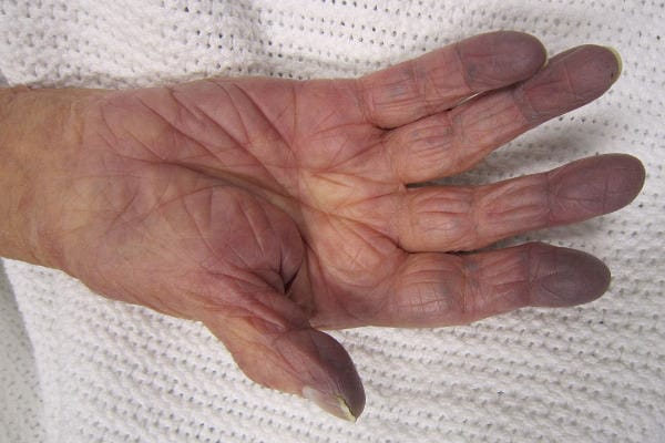
Cyanosis of the fingers
Clinical Diagnosis of Venous TOS
For the most part, doctors can make the presumptive diagnosis of venous thoracic outlet syndrome quite promptly, based on a clinical examination. Most primary care physicians or emergency room physicians will recognize the secondary signs of a venous blood clot, even if they do not directly see the clot. Subsequently, he or she will order imaging tests to confirm the blood clot. Doctors consider the prompt treatment of this clot as the first step in the definitive treatment of venous TOS.
Clinical Diagnosis of Venous TOS
A single large subclavian vein drains most of the blood flow from the arm. Blood clot in the subclavian vein prevents adequate drainage of blood from the arm. In this setting, arm swelling will occur. As a result, patients often describe ‘heaviness’ of the arm. In addition, a blue or purple color may develop, due to loss of oxygen from the trapped blood. In the event the blood clot breaks off, patients may experience a pulmonary embolism. Pulmonary embolism occurs when the blood flow through the lungs is blocked or decreased. In that case, patients will have shortness of breath and chest pain. Pulmonary embolism may be life-threatening.
To begin with, a doctor will look for some or all of the following signs:
Cyanosis is a blue or purple color of the skin. Arterial blood is full of oxygen and appears bright red. Venous blood is low in oxygen and appears dark red to purple. When blood enters the arm but cannot leave normally, more oxygen leaves the blood. As a result, the blood turns darker than normal, and the skin reflects this color change.
Collateral veins appear because the blood cannot leave the arm through the normal venous channels. Blood clot in the large subclavian vein blocks normal blood flow returning towards the heart. As a result, very small superficial veins have to expand to carry the blood back towards the heart. Your doctor can see these collateral veins in the skin of the shoulder and chest. It is important to note that collateral veins represent secondary or indirect evidence of the subclavian vein blood clot.
The venous angle indicates the ability of blood to exit the arm and flow towards the heart. In a normal person, a doctor will slowly raise the arm from the waist until it points forward. In this position, the superficial veins on the back of the hand should empty. If a blood clot is present in the subclavian vein, the doctor will need to raise the arm higher than usual before these veins empty. The venous angle demonstrates secondary or indirect evidence of limited blood flow in the subclavian vein. It may result from blood clot in the vein, or from extrinsic compression of the vein.
Rarely, your doctor may be able to feel the blood clot within the subclavian vein by pressing deep within the armpit.
After a doctor makes the appropriate initial diagnosis of venous TOS, he or she will order imaging tests. The first imaging tests focus on confirming the presence and extent of venous blood clot. Following prompt treatment of this blood clot, further imaging tests evaluate the subclavian vein in detail. These imaging tests focus on demonstrating extrinsic structures compressing the vein and the resulting intrinsic damage to the vein.
Imaging Diagnosis of Venous TOS
When a doctor suspects the diagnosis of venous TOS on clinical examination, he or she will order imaging tests. The imaging diagnosis of venous TOS is quick and accurate. In general, ultrasound, venography, CT venography, or MR venography can confirm the presence of a blood clot. Each of these tests has advantages and disadvantages.
A physician should initially make the presumptive diagnosis of venous TOS based on its dramatic presentation. Typically, one arm appears swollen and blue, and appears quite different from the other arm. In addition, in most cases, the patient notes an abrupt onset of these symptoms. Together with the clinical presentation, the physician can find edema, cyanosis and collateral veins on physical examination. Unfortunately, in most cases, a doctor cannot directly demonstrate the underlying blood clot.
Therefore, after the physician makes the initial diagnosis of venous TOS, imaging diagnosis of venous TOS serves several purposes. Firstly, imaging tests confirm the presence and extent of venous blood clot. Secondly, imaging tests clarify the extrinsic structures compressing the vein. Thirdly, imaging tests show the intrinsic damage to the vein. It is important to realize that arm motion forwards or over the head causes vein compression in many people. Therefore, experts perform imaging tests with the arms down and with the arms elevated.
Some specialists have suggested that hypercoagulable blood, or ‘thick’ blood that clots easily, causes venous TOS. For this reason, some physicians will obtain blood clotting tests in patients with venous TOS. At this time, there is little data to support routine testing of blood clotting factors. However, in patients with other clinical evidence of a blood clotting disorder, the physician may consider blood testing.
It is important to bear in mind that venous TOS frequently recurs following treatment. Therefore, many physicians perform routine follow-up imaging for early detection of recurrent stenosis or blood clot.
Imaging diagnosis of venous TOS
While there are several tests that doctors use to diagnose venous TOS, each has its advantages and disadvantages. Treatment of venous TOS has evolved significantly, although debate continues regarding optimal treatment methods and timing. Nonetheless, if a doctor diagnoses and treats venous TOS promptly, treatment outcomes are usually excellent.
 X-rays show excellent bone detail, but do not show soft tissue detail, and are limited to two dimensions. X-rays do not show arteries or veins, and are almost always normal in patients with venous TOS. In addition, venous TOS is likely to occur without cervical rib or other bone anomaly. Therefore, x-rays have no significant diagnostic or therapeutic value in patients with venous TOS.
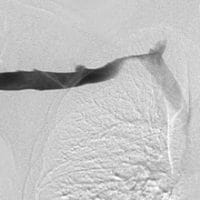 Direct venography produces excellent images of the subclavian vein on one side. In detail, the physician inserts a small catheter into a hand or arm vein and injects dye into the vein. During the injection, the physician takes X-ray images as the dye passes through the veins towards the heart. Direct venography provides two major advantages besides confirming the presence of a clot. Firstly, a physician can perform thrombolysis, injection of a clot-dissolving medicine, directly after the venogram. Additionally, the physician can perform venoplasty, dilating a venous stenosis with a balloon, directly after the venogram. Notably, direct venography can examine only one side at a time, and utilizes a moderate radiation dose. As a result, a physician would need to perform two separate procedures in patients with bilateral symptoms. Significantly, direct venography can provide excellent information in patients with surgically-altered anatomy, including dialysis fistula or graft. CT venography or MR venography demonstrate structures that cause compression of the vein, which can be valuable for planning surgery.
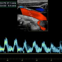 Ultrasound is performed without any radiation, and demonstrates blood flow in the veins in real time. This allows ultrasound evaluation during dynamic movement of the arms. At the same time, bones interfere with ultrasound and create blind spots. Thus, ultrasound may not directly demonstrate venous compression in the thoracic outlet. However, ultrasound shows blood clots quite well, as well as abnormal venous waveforms just beyond the blind spot. Ultrasound does not demonstrate fibrous bands and muscle anomalies in detail.
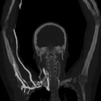 CT venography (CTV) is performed by injecting contrast into a peripheral vein, without an arterial catheter. CTA is fast, and shows excellent detail of bones and arteries. However, CT venography does not show soft tissues in detail, including fibrous bands and muscle anomalies. Additionally, the patient receives a significant radiation dose during a CTV. In many centers, CTV is the first-line test for many vascular surgeons.
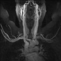 MR venography (MRV) can be performed in two ways. First is direct MRV. The physician injects contrast into a small arm vein. Shortly thereafter, the MRI scanner creates images as the contrast passes through the arm and chest. Direct MRV shows only veins, and only in one arm. In contrast is indirect MRV. Again, the physician injects contrast into a small arm vein. In contrast to direct MRV, scanning begins after a delay. First, injected contrast travels through the heart, the lungs, the arms, and returns through the arm veins. At this time, the MRI scanner creates images, showing both arteries and veins, in both arms. MRV shows the veins while eliminating other structures. MRV requires no radiation. If used in conjunction with MRI, superb soft tissue detail is available. Excellent depiction of fibrous bands, muscle anomalies, bones, and brachial plexus is possible with the combination of MRV and MRI.
Interactive Media
This patient had an MR angiogram, followed by an MR venogram. Slide the handle side to side to see the difference between the two studies.

Diagnosis of Venous TOS
Doctors usually suspect the diagnosis of venous TOS early in the course of the disease. In the first place, venous TOS usually presents in a quite dramatic fashion. A doctor is not likely to miss this presentation. In general, doctors begin treatment promptly after confirming the diagnosis of venous TOS.
Symptoms
Venous TOS occurs as a result of a blood clot in the large subclavian vein that drains the arm. Blood enters the arm, but cannot easily leave. Thus, patients notice swelling, heaviness, and abnormal color of the affected arm. Symptoms may arise slowly or suddenly. Rarely, blood clots travel to the lungs, and the patient may develop shortness of breath.
Clinical Diagnosis
The doctor will notice a difference in size between the two arms. Change in color of the skin may appear as a result of slow blood flow and loss of oxygen. The doctor may notice additional abnormal veins of the arm, shoulder or chest wall.
Laboratory Diagnosis
Some authorities believe that abnormal clotting of the blood contributes to some cases of venous TOS. For this reason, some doctors include laboratory assessment of blood clotting when evaluating a patient with venous TOS. However, in most cases, laboratory tests do not contribute to the diagnosis or treatment of venous TOS.
Imaging Diagnosis
Imaging tests provide valuable diagnostic information in patients with venous TOS. In addition, much of the information provided by imaging tests will guide treatment. With this in mind, it is helpful to understand the advantages and disadvantages of the available imaging tests.
Summary: Diagnosis of Venous TOS
Doctors usually find the diagnosis of venous TOS straightforward and obvious. It is important to understand the symptoms, physical examination, lab tests and imaging tests that contribute to the diagnosis. In particular, imaging tests confirm the diagnosis of venous TOS, but also provide information that is vital to treatment planning. Overall, it is important to diagnose and treat the venous blood clot promptly. If you suspect you have the diagnosis of venous TOS, you should promptly find a physician to confirm the diagnosis.
