

Types of Thoracic Outlet Syndrome
Types of Thoracic Outlet Syndrome
Table of Contents
Table of Contents
The Classical Description: 3 Types of Thoracic Outlet Syndrome
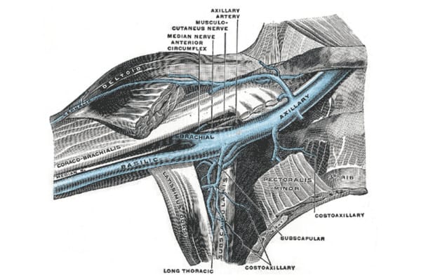
Thoracic outlet syndrome specialists classically define three types of thoracic outlet syndrome. In this framework, each type of thoracic outlet syndrome gets defined by the primary structure that is affected. Specifically, the three primary structures that pass through each thoracic outlet are an artery, a vein and a nerve plexus. Thus, the three classic types of thoracic outlet syndrome are arterial thoracic outlet syndrome, venous thoracic outlet syndrome, and neurogenic thoracic outlet syndrome.
To clarify, each of the three types of TOS is defined by the vital structure that is compressed in the thoracic outlet:
- Compression of the brachial plexus: Neurogenic thoracic outlet syndrome
- Compression of the subclavian vein: Venous thoracic outlet syndrome
- Compression of the subclavian artery: Arterial thoracic outlet syndrome

For the purposes of learning about each of the three major types of thoracic outlet syndrome, we will discuss each type of thoracic outlet syndrome separately. However, compression of one vital structure may include coincident compression of the adjacent vital structures. For this reason, there may be clinical signs of more than one type of thoracic outlet syndrome in any patient. The NeoVista® MRI examination proves extremely valuable in many of these cases.
What are the 3 Types of Thoracic Outlet Syndrome?
- Neurogenic Thoracic Outlet Syndrome
- Venous Thoracic Outlet Syndrome
- Arterial Thoracic Outlet Syndrome
Neurogenic Thoracic Outlet Syndrome
Neurogenic thoracic outlet syndrome is by far the most common type of thoracic outlet syndrome. At the same time, neurogenic TOS is the most difficult type of thoracic outlet syndrome to diagnose. Patients often cycle through many doctors, and many specialities, before they receive the correct diagnosis. And, unfortunately, in many cases, doctors may not even make the correct diagnosis. So patients with neurogenic TOS often suffer needlessly for years.
In general, compression of a single nerve anywhere in the body may cause symptoms such as pain, numbness, tingling and weakness. Doctors describe this process as a nerve entrapment syndrome. Neurogenic thoracic outlet syndrome occurs through the same process, but is much more complex than entrapment of a single peripheral nerve.
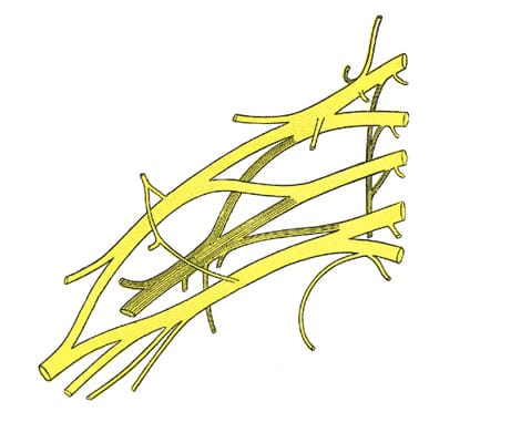
In detail, five nerve roots arise from the spinal cord on each side of the neck. After leaving the spinal cord, these nerve roots enter the thoracic outlet. In each thoracic outlet, the nerve roots form a complex branching network, called the brachial plexus. Compression or tension on any part of the brachial plexus may cause symptoms, resulting in neurogenic thoracic outlet syndrome. Given the complexity of the structure of the brachial plexus, patients can suffer a broad range of symptoms.
Thus, doctors see a complex clinical presentation, since compression may involve any number or combination of the branches of the brachial plexus. Symptoms may include pain, numbness, tingling, coldness and weakness of the affected upper extremity. The neurogenic type of TOS may cause symptoms in one or both arms or hands, as well as the neck, chest, back or shoulders.
Venous Thoracic Outlet Syndrome
Venous TOS is an uncommon type of TOS. Venous TOS occurs when a blood clot forms in the large vein that drains the arm. Doctors should suspect this type of TOS when a patient presents with arm swelling and color changes. Many available diagnostic tests can easily demonstrate the blood clot. Treatment of this type of TOS is urgent, and doctors usually strive to dissolve the blood clot quickly.
A single, large subclavian vein drains the blood from each arm. Compression of this vein can prevent normal venous drainage of the arm. When compression is repetitive, severe, or prolonged, it can cause damage to the inner wall of the vein. As a result of this damage, blood clot can form within the vein. In this event, swelling, heaviness and cyanosis (abnormal blue color) develops in the affected arm. Some patients may develop new superficial veins of the chest and shoulder, as blood must return to the heart through new pathways.
When a doctor sees a patient with this clinical picture, they should immediately consider the diagnosis of venous TOS. This type of TOS accounts for about 2 to 5% of all TOS cases. Quick and accurate diagnosis is vital to prevent complications such as blood clot fragments traveling to the lungs or, rarely, to the brain, causing a stroke.
Treatment of venous thoracic outlet syndrome should begin urgently, using methods to dissolve or remove the blood clot in the subclavian vein. After this, imaging studies can show whether the vein is damaged or compressed by external structures. If these structures are not removed or reduced, the same damage and blood clot may recur, resulting in the same type of TOS.
Arterial Thoracic Outlet Syndrome

Arterial TOS is the rarest type of TOS. This type of TOS occurs when compression of the subclavian artery causes arterial damage, with secondary blood clot causing symptoms. Blood clot forms in the damaged arterial segment, and fragments travel down the arm, creating the sudden severe blockage of blood flow. Doctors typically recognize arterial thoracic outlet syndrome quickly, and treat it by removing the clot emergently.
Arterial TOS develops after compression of the subclavian artery causes arterial damage or blood clot. The subclavian artery provides blood flow to each arm. Repetitive and severe compression of this artery in the thoracic outlet causes damage to the arterial wall. Damage limited to the inner arterial wall can cause a fixed scar and stenosis (narrowing of the artery). In contrast, damage to the full thickness of the wall can result in an aneurysm (focal ballooning of the artery). Arterial blood clots may form in the damaged arterial segment. Blood clots can break off and travel down the arm and block distal arteries, causing sudden and severe loss of blood flow. Doctors call this process acute arterial insufficiency.
Arterial TOS accounts for about 1% of all TOS cases. Most patients with this arterial TOS present with an acute onset of symptoms, including pain and loss of pulses. It is important to realize that if not treated emergently, loss of tissue (gangrene) may occur. Therefore, doctors need to recognize and treat this arterial TOS emergently. Initial treatment requires emergent removal or dissolving of the arterial blood clot. Imaging tests can then show the damaged arterial segment and the external structures that caused the damage. Later, doctors typically repair the damaged arterial segment and remove or release the abnormal external structures causing compression of the artery.
Do you have pain, numbness, tingling or weakness? Click to learn more about the diagnosis of thoracic outlet syndrome.
Learn how the NeoVista® MRI for TOS provides diagnostic information that is not available elsewhere.
Best treatment of thoracic outlet syndrome requires a dedicated team of thoracic outlet syndrome specialists.


