

Diagnosis of Neurogenic Thoracic Outlet Syndrome
Diagnosis of Neurogenic Thoracic Outlet Syndrome

The accurate and timely diagnosis of neurogenic thoracic outlet syndrome (TOS) is critically important in determining the outcome of the disease. Since arterial and venous thoracic outlet syndrome often have dramatic presentations, doctors usually confirm those diagnoses promptly. This early diagnosis enables definitive surgical treatment early in the course of the disease. On the other hand, the diagnosis of neurogenic TOS often has a more insidious and intermittent presentation. Thus, the accurate and early diagnosis of neurogenic thoracic outlet syndrome may be elusive. However, accurate diagnosis and early treatment are important in shortening the course of the disease and in preventing permanent pain or motor function loss. Read more about the unique diagnosis of neurogenic thoracic outlet syndrome.
Table of Contents
Table of Contents
Symptoms of Neurogenic Thoracic Outlet Syndrome
Neurogenic TOS is the most common form of thoracic outlet syndrome. At the same time, the diagnosis of neurogenic thoracic outlet syndrome challenges physicians much more than the diagnosis of venous TOS or arterial TOS. Symptoms of neurogenic TOS are often non-specific and confusing. A doctor may find a completely normal neurological examination if he doesn’t attempt specific tests for neurogenic TOS. Equally important, patients may not understand the confusing symptoms of neurogenic TOS that might cause them to seek a TOS specialist.

Compression, tension, and resultant damage of the nerves of the brachial plexus cause the symptoms of neurogenic TOS. However, the brachial plexus is quite complex, comprising different nerve fibers, with multiple branches and divisions. Compression of different fibers or branches will cause different symptoms. Additionally, compression may only occur in certain positions of the arms and neck. As a result, patients may fail to understand why they are experiencing pain at certain times, in certain occupations or recreations. Nonetheless, patients suffer considerable pain of different types and causes.
Patients with neurogenic TOS may experience one or more of the following symptoms of the chest, arm, wrist, or hand:
Pain Mechanisms
In general, doctors divide the mechanisms of pain into two broad categories: nociceptive pain and neuropathic pain.
Firstly, it is helpful to understand the normal pain pathway. Peripheral receptors of various types are located throughout the body. A peripheral receptor might be specialized for pressure, pain, position or temperature. A peripheral nerve typically connects peripheral receptors to the brain. When an appropriate stimulus triggers one of these receptors, the peripheral nerve transmits a signal from the receptor to the spinal cord, where it usually connects to another nerve, and then to the brain.
Second, we can broadly define the two basic mechanisms of pain:
- Nociceptive pain-A normal nerve transmits a pain signal from a normal peripheral receptor to the brain from a site of tissue damage, such as a burn or a cut in the skin.
- Neuropathic pain-A peripheral nerve is damaged somewhere along its course between the peripheral receptor and the brain. The peripheral receptor and overlying tissue are normal. In this case, the brain receives a signal from the abnormal, damaged peripheral nerve, but interprets the signal as coming from the peripheral receptors.
Third, neuropathic pain is a clinical descriptor, but not a specific diagnosis. Bear in mind that pain syndromes are complex. There are pain syndromes that are incompletely understood, and do not fall neatly into these two categories. Complex Regional Pain Syndrome (CRPS) represents one example of such a condition. Neuropathic pain may result from chronic diseases that affect the peripheral nerves, such as diabetes. Additionally, neuropathic pain may be acute or chronic. It can arise from the peripheral nerves, but also from the central nervous system (the brain and spinal cord).
Neurogenic TOS represents one example of neuropathic pain. The peripheral tissues and receptors are normal. However, entrapment, compression, and tension of the brachial plexus causes damage to its component nerves. These nerves send abnormal signals to the brain.
All of the different types of peripheral receptors of the arms send peripheral nerve fibers through the brachial plexus. The brachial plexus has a complex branching pattern. Given these facts, one can understand the complexity of symptoms that neurogenic TOS patients experience.
Course of Symptoms
The symptoms of neurogenic TOS may develop suddenly or gradually. Some patients develop the symptoms of neurogenic TOS suddenly following a motor vehicle accident. Others develop symptoms after longer periods spent at repetitive occupations. In particular, computer users carry significant risk for developing neurogenic TOS. Other occupations requiring physical activity may predispose workers to developing neurogenic TOS.
Symptoms of neurogenic TOS may occur in a progressive or intermittent fashion. Some patients may experience symptoms after a long work day, with symptoms resolving at night. If the disease is not diagnosed and treated, these patients may notice symptoms lasting into the evening or overnight. Eventually, symptoms may persist over the weekend. In other patients, the symptoms of neurogenic TOS begin, never resolve, even briefly, and may worsen and progress.
Additionally, symptoms may depend upon position and posture. Some patients are able to relieve or reduce their symptoms with certain arm or neck positions. Others find relief when laying down or sitting in a certain position. Unfortunately, symptoms often progress to the point where these measures fail to help.
Are these symptoms unique to thoracic outlet syndrome?
Unfortunately, no, many symptoms of neurogenic TOS occur in other diseases. Patients with disease of the cervical spine (neck), such as disk herniations, may have similar symptoms. Other upper extremity nerve entrapment syndromes, such as carpal tunnel syndrome, may behave in a similar fashion. Nerve diseases such as complex regional pain syndrome (CRPS), may be confused with neurogenic TOS. An experienced TOS specialist is the best doctor to begin distinguishing these symptoms and their underlying causes. In many cases, advanced imaging provides critical information to confirm the diagnosis.
Clinical Diagnosis of Thoracic Outlet Syndrome
The clinical diagnosis of neurogenic thoracic outlet syndrome challenges physicians. Patients with neurogenic thoracic outlet syndrome experience a broad range of symptoms that appear confusing to many doctors. In addition, many doctors may not know the best thoracic outlet syndrome test. Fortunately, an experienced thoracic outlet syndrome specialist will recognize the pattern of symptoms of neurogenic thoracic outlet syndrome symptoms. In many cases, your primary care physician may be the first person to make the diagnosis of neurogenic thoracic outlet syndrome. Because these symptoms are non-specific, your TOS specialist should know how to order the most accurate and helpful thoracic outlet syndrome tests. Experienced, dedicated thoracic outlet syndrome imaging remains a preferred test of many TOS specialists.

An experienced TOS specialist focuses the clinical diagnosis of neurogenic thoracic outlet syndrome on two important components: an accurate and detailed medical history and a specialized physical examination.
Medical History
In general, a doctor evaluates a patient by taking a medical history. In detail, the medical history starts with the ‘presenting complaint’ or chief complaint. The chief complaint comprises the primary symptom causing the patient to visit a doctor. After hearing the patient’s chief complaint, a doctor gathers details by taking a history of present illness. Typically, the following details make up the history of present illness:
History of Present Illness
History of Present Illness Related to Neurogenic Thoracic Outlet Syndrome
Moreover, an experienced TOS specialist will zero in on details that are specific to neurogenic TOS:
Past Medical History Related to Neurogenic Thoracic Outlet Syndrome
Besides a detailed and specific thoracic outlet syndrome history of present illness, a thoracic outlet syndrome specialist would investigate possible causative factors of neurogenic TOS. Specifically, any history of overuse, trauma, and occupational or recreational stressors could contribute to the development of neurogenic TOS. Additionally, an experienced TOS specialist would fully explore the time course of neurogenic symptoms.
For example, many TOS patients are able to define a specific traumatic episode that created immediate symptoms. Notably, many TOS patients have suffered a neck injury in a motor vehicle accident. Altrnatively, a thoracic outlet syndrome patient may relate a work-related injury, while other TOS patients describe a recreational injury. In particular, some TOS patients note sudden onset of symptoms after activities like rock climbing or weightlifting.
Nonetheless, while post-traumatic neurogenic TOS frequently develops with a sudden onset, some trauma patients experience a delayed or gradual onset.
In contrast, patients who develop neurogenic TOS from overuse frequently cannot define a specific event or time when they first experienced symptoms. These patients often describe symptoms that were so mild as to be of little concern. Yet over time, the symptoms worsened to the degree that they impacted the patient’s lifestyle. These patients often relate a history of mild symptoms occurring after long episodes of recreational or occupational overuse. These symptoms would spontaneously resolve after the activity. Over time, if the inciting activity continued, symptoms would persist for hours, and eventually overnight. Even at this point, patients frequently would note relief over weekends. However, even these respites would fade, and symptoms would persist from that point forward.
Work and Lifestyle History Related to Neurogenic Thoracic Outlet Syndrome
Following the history of present illness, a TOS specialist would expand other elements of the patient’s overall medical history that are pertinent to neurogenic TOS.
Past Medical History
Has the patient ever broken his or her collarbone or ribs? Has the patient had surgery to the cervical spine, neck, chest, or shoulder? What drugs has the patient taken in the past, or is currently using? What treatments has the patient previously tried for TOS, such as physical therapy or acupuncture?
Occupational History
Does the patient’s occupation require physical activity, or does it demand unusual posture or prolonged posture? Does the occupation demand a lot of arm use, or positioning of the arms in unusual positions? Does the patient use a computer? Is there lifting involved in work, particularly with the arms forward or overhead?
Recreational History
What sports does the patient participate in? Do these involve use of the arms overhead? Does the patient use computers, phones or tablets on a regular basis?
Other History Pertinent to Neurogenic Thoracic Outlet Syndrome
In what position does the patient sleep? Does the patient carry young children? Has the patient ever experienced a blood clot?
Physical Examination
It is important to realize that the standard neurological examination is insufficient for the diagnosis of neurogenic thoracic outlet syndrome. When a patient presents to a physician with arm and neck pain, the physician typically performs a standard neurologic examination. Unfortunately, this examination would be unlikely to rule in or rule out neurogenic TOS.
For this reason, doctors must be familiar with the specialized physical examination tests that can demonstrate findings of neurogenic thoracic outlet syndrome. A standard neurological examination is not adequate as a thoracic outlet syndrome test. Instead, an experienced thoracic outlet syndrome specialist performs a series of specialized provocative thoracic outlet syndrome tests. These tests are intended to demonstrate compression of blood vessels or nerves, or tension on nerves, in the thoracic outlet.
The diagnosis of neurogenic thoracic outlet syndrome has evolved over a long period of time. TOS was first described in 1818. Since then, experts have studied the anatomy and causes of TOS. Many of these experts have devised provocative tests to diagnose thoracic outlet syndrome quickly and accurately. Notably, most of these tests have been based on changes in blood flow, rather than nerve compression or tension. At the same time, most patients with TOS have neurogenic TOS, making tests of blood flow quite limited for diagnosis. More recent peer-reviewed evaluation of these provocative these tests has proven that they have limited sensitivity, specificity, or accuracy.
It should be noted that compression of arteries or veins does not confirm compression of nerves. Before the invention of modern medical imaging, doctors could not distinguish compression of blood vessels from compression of nerves with any confidence. As a result, past experts used tests of vascular compression as proxies for nerve compression, since they had no better test. Obviously, we now have better tools, in particular imaging tests, to distinguish compression of blood vessels and nerves. Unfortunately, some clinicians still use tests of blood flow in patients with nerve compression.
Experienced TOS specialists realized that they needed to improve the clinical evaluation of nerve compression. These doctors developed specialized thoracic outlet syndrome tests specifically to evaluate nerve compression. Knowledgeable TOS specialists use these specialized tests today. It is important to realize that while specific tests for nerve compression are better than tests of blood flow, they are not highly accurate, and do not distinguish the underlying anatomic causes of neurogenic TOS. Instead, they provide additional evidence for the diagnosis of thoracic outlet syndrome in a patient with a suitable clinical presentation and corresponding signs and symptoms. Finally, while tests of blood flow may overlap those for nerve compression, TOS specialists understand how to distinguish these results.
These tests are called ‘provocative’ tests. To explain, the examining physician moves the patient’s arms or neck into specific positions during each test. These positions temporarily increase compression of blood vessels or nerves in order to reproduce the patient’s symptoms and signs.
Adson’s Test
The patient assumes a sitting position, with arms at the side. The patient turns his head towards the affected side, extends his neck (bends his neck backwards), and takes and holds a deep inspiration. The physician monitors the patient’s radial pulse at the wrist. If the pulse diminishes or disappears, or if the patient’s symptoms are reproduced, Adson’s test is positive.
A variant of Adson’s test is the reverse Adson’s test, where the procedure is performed in identical manner, but with the patient’s head turned away from the affected side.

Halstead Maneuver
With the patient sitting, the examiner puts his or her fingers on the radial pulse at the wrist. After the pulse is localized, the examiner puts downward traction on the arm, rotates the arm in the outward direction, and moves the arm back and away by about 45 degrees. In some descriptions, the patient turns their head to the opposite side during the test. The Halstead maneuver is considered positive if the radial pulse decreases or disappears.
Presumably, the maneuver causes compression of the subclavian artery in the costoclavicular interval. In the past, compression of the artery was thought to correlate with compression of the brachial plexus. However, peer-reviewed medical articles do not support this theory. Also, in our imaging experience, compression of the artery occurs in isolation in some patients, but with compression of the brachial plexus in other patients. The degree of brachial plexus compression in this setting ranges from none to severe. Therefore, like all physical examination tests that rely on the radial pulse, the Halstead maneuver should not be considered diagnostic of neurogenic TOS.

Wright Test
The patient is positioned sitting or supine (lying on her back). The examiner lifts the patient’s arm overhead to an angle greater than 90 degrees at the shoulder, with the elbow straight or bent less than 45 degrees, while monitoring the patient’s radial pulse. A decreased or absent radial pulse, or reproduction of the patient’s symptoms, is a positive test, and is thought to be caused by narrowing of the costoclavicular interval.
Novak and McKinnon have adapted this test by keeping the elbow extended and the wrist in neutral as not to provoke cubital tunnel syndrome (entrapment of the ulnar nerve at the elbow) or carpal tunnel syndrome (entrapment of the median nerve at the wrist).
https://youtu.be/L6BoVyE_vfE?t=10s
Roos’ Test
The patient can be sitting or standing for the Roos’ test. The patient holds their arm in the “stick ‘em up” position, with the shoulders raised 90 degrees, the elbows bent 90 degrees, and the hands above the head, with palms facing forward and the head and neck in neutral position. The patient then opens and closes their hands for three minutes. A positive test occurs when the patient’s symptoms are reproduced within three minutes.
The Roos’ test was conceived by Dr. David Roos, a pioneering surgeon in the 1970’s who catalogued many of the soft tissue abnormalities of the thoracic outlet in patients with TOS. The Roos’ test is also known as the EAST test (Elevated Arm Stress Test) or Abduction External Rotation Test (AER Test).


Costoclavicular Test (Eden Test)
The patient is sitting, and assumes a ‘military’ posture, with the back straight and the shoulders pushed backward and downward. The physician may help depress the shoulder on the side being examined, while monitoring the radial pulse at the wrist. If this pulse decreases or disappears, or if the patient’s symptoms are reproduced, the test is positive. A positive test is assumed to be due to narrowing at the costoclavicular interval, causing compression of the subclavian artery or brachial plexus. Importantly, reduction or loss of the radial pulse without neurogenic symptoms should not be interpreted as evidence of neurogenic thoracic outlet syndrome.
https://www.youtube.com/watch?v=Mifo6ZzkNLA
Tinel’s Sign
This is also known as pressure provocation testing. Tapping with the examiner’s finger over a nerve may produce a tingling sensation within the distribution of that nerve. The test is performed over common entrapment sites in the upper extremity, such as the carpal tunnel, the cubital tunnel, and the brachial plexus in the supraclavicular fossa. This test tends to be positive in later stages of chronic nerve compression.

Upper Limb Neural Tension Test
Butler (1) described upper limb neural tension tests. A thoracic outlet syndrome specialist performs these tests by placing the patient n a sitting position followed by gradual repositioning of the joints of the upper extremity. The specialist starts by raising the shoulder away from the body with the elbow bent, then straightening the elbow, and then moving the wrist and forearm into various positions. The test is intended to place the nerves that stretch from the spinal cord to the fingers under tension. The test is considered positive when it reproduces neurogenic symptoms.
Although Butler devised several variants of this test, he emphasized the upper limb nerve tension test 3 (ULTT 3). This variant is more sensitive for ulnar nerve irritation. Butler also recommended performance of the slump test in longsitting and sitting to investigate the spinal canal components for adverse neural tension. He also suggested that the possibility of double or multiple crush syndromes should be investigated, as well as the contribution of the sympathetic nervous system to the symptoms experienced by some patients, such as color or temperature changes in the hands.


Spurling’s Test
Most patients with neurogenic TOS have neck and arm pain. Pain may arise from compression or stretching of the nerves in the spine or in the thoracic outlet. A knowledgeable specialist will attempt to rule out narrowing of the cervical spine through Spurling’s test. Spurling’s test can suggest entrapment of the nerve roots as they exit the spinal canal. It should be remembered that most patients with neurogenic TOS are young and otherwise healthy. Thus, these patients are unlikely to have chronic degenerative changes or narrowing of the cervical spine. For this reason tests of the cervical spine should be performed, but are typically negative.

Can a patient have multiple types of thoracic outlet?
Yes, one patient can have multiple types of thoracic outlet syndrome at the same time!
As we know, the subclavian artery, subclavian vein, and brachial plexus pass through each thoracic outlet in close proximity. Furthermore, the mechanisms of compression are relatively similar for all types of thoracic outlet syndrome. Thus, a patient with thoracic outlet syndrome can experience compression of 1, 2, or all 3 of these vital structures. In fact, many patients with neurogenic thoracic outlet syndrome have significant compression of the subclavian vein, but without a blood clot. Since this compression impairs venous drainage of the arm, swelling or edema of the arm can result. As a result, symptoms of neurogenic thoracic outlet syndrome may be magnified. In that case, treatment to relieve the venous compression helps relieve neurogenic thoracic outlet syndrome.
The clinical diagnosis of thoracic outlet syndrome may fail to distinguish these multiple compression effects. Therefore, modern imaging like our NeoVista® MRI examination is a highly valuable tool to provide this important additional information prior to treatment.
Watch videos about the clinical diagnosis of thoracic outlet syndrome on the TOS MRI YouTube channel or toseducation.org
Imaging Diagnosis of Neurogenic TOS
Modern imaging provides essential information in patients with neurogenic TOS. Firstly, imaging tests can directly or indirectly demonstrate compression of veins, arteries or nerves. Second, imaging tests can prove secondary damage to these structures, such as an aneurysm. Third, imaging tests can rule in or rule out blood clots, which are a critical factor in determining treatment. Fourth, imaging tests show multiple points of compression that affect treatment differently. Finally, imaging thoracic outlet syndrome tests rule out abnormalities in the cervical spine, chest, or shoulder.
Clinical tests for neurogenic TOS demonstrate limited sensitivity and specificity. We know that the standard neurological examination often misses the diagnosis of neurogenic TOS. Even specialized provocative TOS tests have proven to be limited in their ability to diagnose or rule out TOS. Additionally, even when these clinical tests are accurate, they do not demonstrate the anatomic or pathologic abnormalities that are causing compression. In contrast, imaging tests demonstrate the anatomy and pathology in patients with TOS in great detail.

Different imaging tests have advantages and disadvantages. X-rays are inexpensive and fast, but do not show crucial soft tissues. Ultrasound is widely-available, and shows soft tissues and blood flow in real time, but is blocked by bones and lung. Direct angiograms and venograms are invasive, and do not show crucial soft tissues outside the blood vessel. CT scans are fast and detailed and show great bone detail, but are limited for soft tissues and require higher radiation doses. MRI scans show great soft tissue detail, require no radiation, and can show blood vessels and nerves, but bone detail is limited. An experienced thoracic outlet syndrome specialist will know which test to apply for which form of TOS.
Treatment decisions can change according to the results of imaging tests. Different mechanisms cause different types of TOS, and multiple mechanisms can exist in one patient. Doctors can start conservative treatment in one patient, or surgical treatment in another, based on these findings. Additionally, different methods of conservative treatment and different types of surgery may be suggested by imaging findings. Given the anatomic variations in the thoracic outlet, multiple different points of compression are possible. It is hard to imagine that one type of conservative treatment, or one surgery, can correct the unique problem without knowing the detailed anatomy and pathology provided by imaging.
The NeoVista® MRI study gives the patient and doctor unique and valuable information. NeoVista® includes separate and detailed images of the soft tissues, nerves, and blood vessels in one comprehensive study. NeoVista® demonstrates changes on arm motion. And NeoVista® is performed by people with world-class knowledge, earned through years of study, experience, and motivation.
Imaging diagnosis of neurogenic TOS
Imaging of patients with neurogenic TOS provides valuable information to the TOS specialist. While a TOS specialist may strongly suspect that his or her patient has neurogenic TOS, the clinical examination has poor sensitivity and accuracy. Therefore, several outstanding questions must be answered. Firstly, can compression or tension of the brachial plexus be proven? Second, is there intrinsic disease of the brachial plexus? Third, can alternative diagnoses, such as a herniated disc, be ruled out? Fourth, what are the exact anatomic structures that cause compression or tension of the brachial plexus? Fifth, does arm motion contribute to compression? Finally, what other structures are involved, such as arteries or veins?
Fortunately, a dedicated TOS MRI can answer all of these questions and more. Done properly and interpreted by a TOS-specialized radiologist, MRI provides extremely valuable information to the TOS specialist.
What would the ideal imaging test for neurogenic thoracic outlet syndrome show?
How does a TOS specialist use this imaging data?
It is important to remember that clinical tests used by TOS specialists do not possess very good accuracy. Therefore, while a TOS specialist may suspect neurogenic TOS, he or she cannot easily prove the diagnosis. So, MRI proof of brachial plexus compression or tension provides critical data. Also, rarely, inflammation or trauma of the brachial plexus may cause a similar clinical picture as neurogenic TOS. MRI can clearly rule out such brachial plexus damage.
TOS specialists often find a challenge in distinguishing the symptoms of neurogenic TOS from those caused by cervical spine disease. The NeoVista® MRI examination always includes images of the cervical spine, just for this purpose.
Once the diagnosis of neurogenic TOS is confirmed, treatment decisions are based on the underlying cause. Patients with extra or abnormal ribs may require surgery. On the other hand, patients with soft tissue abnormalities or abnormalities of shoulder motion can respond quite well to conservative treatment. Additionally, given the anatomic complexity of the thoracic outlet, MRI can help surgeons determine their surgical goals. In most surgical specialties, surgeons have refined and minimized their procedures over many decades. Many surgeons intend to resolve pathologic structures while minimizing their effect on normal structures. MRI is widely used in other surgical fields for this purpose. In a similar fashion, MRI may be used in TOS patients for the same reason.
Most neurogenic TOS patients experience symptoms or worsen their symptoms when performing activities. If these changes can be evaluated, conservative treatment plans can be tailored to a specific patient. The NeoVista® MRI uses a proprietary quantitative method of evaluating arm motion on each side, in each patient. We believe this data adds a level of detail that a TOS specialist would find quite important and helpful.
The same process that damages the brachial plexus may compress or damage arteries or veins. Should such compression or damage exist, the surgical plan might require a second, specialist surgeon. MRI provides this data before surgery, which could change the surgical plan.
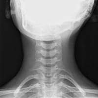 X-rays show good bone detail, but do not show soft tissue detail, and they are limited to two dimensions. X-rays do not show the brachial plexus, and are almost always normal in patients with neurogenic TOS. In addition, the presence of cervical ribs does not increase the likelihood of neurogenic TOS in any single patient. Therefore, x-rays hold little diagnostic or therapeutic value in patients with neurogenic TOS.
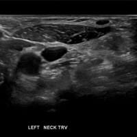 Ultrasound is performed without any radiation, and can distinguish fat, muscle, blood vessels and nerves. Additionally, ultrasound can demonstrate real-time dynamic movement of the arms. At the same time, bones interfere with ultrasound and create blind spots. Thus, ultrasound cannot demonstrate compression of the brachial plexus in the costoclavicular interval, which occurs commonly in these patients. Finally, ultrasound does not clarify fibrous bands and muscle anomalies in detail, which commonly cause brachial plexus compression.
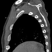 CT, also known as ‘CAT scan,’ utilizes a spinning x-ray generator to create images in 3 dimensions. CT is widely available, and fast, and it shows excellent bone detail. CT images can be reformatted into 3-dimensional models. While CT can easily prove narrowing of the costoclavicular interval, it does not show the brachial plexus well. Additionally, CT does not show other soft tissues well, including fibrous bands and muscle anomalies that frequently compress the brachial plexus. Finally, the patient receives a significant radiation dose during a CT scan. In many centers, CT is the first-line test for patients with neurogenic TOS.
 MRI can show the soft tissues of each thoracic outlet in great detail. Scalene muscles and their anomalies, fibrous bands, the brachial plexus and its branches, and the fat that surrounds these structures are very well seen. Bones such as the clavicle, scapula, first rib, cervical rib, and cervical spine can also be seen. MRI can be performed with the arms in different positions. Radiologists can see the changes that arm motion causes.
Where can you find an imaging test that answers these questions?
Obviously, we believe in the value of MRI as the best imaging tool for patients with neurogenic TOS. Further, the test and interpretation of this complex disease should be placed in the hands of experienced TOS specialists. At the risk of sounding biased, we feel strongly that the NeoVista® MRI examination answers each of the important questions in patients with neurogenic TOS, and provides experienced TOS expertise.
The full NeoVista® MRI examination is completed in one visit. It includes detailed images and expert interpretation, including the soft tissues and bones of each thoracic outlet, the cervical spine, the brachial plexus, and the subclavian artery and vein. Arm motion is evaluated and measured. This evaluation can be repeated on follow-up examination. In addition, we support your TOS specialist by offering review and discussion of each case. In many cases, we can provide a video teleconference with your specialist and you, including review of your images.
You can try this simple example that shows one component of our test. Grab the handle and slide back and forth to see two different levels of detail of the brachial plexus.
Interactive Media
MRI can demonstrate the brachial plexus in detail. A radiologist can zoom in to evaluate the branching pattern, size, brightness, and course of each nerve root in the brachial plexus.


MRI has revolutionized the diagnosis of neurogenic thoracic outlet syndrome
In 1895, Conrad Wilhelm Roentgen produced the first medical X-ray image. Since then, medical imaging has revolutionized the practice of medicine. Especially over the last few decades, advances in computer technology have led to advances in medical imaging. CT, ultrasound, PET, and MRI are now widely available. Doctors use such imaging tests millions of times every year. These imaging tests help doctors make diagnoses and treatment decisions every day.
And now we can apply the same technology to the challenge of TOS.

What are the symptoms of TOS?

Which clinical tests are used to diagnose TOS?

Are lab tests useful for the diagnosis of TOS?

What imaging tests accurately diagnose TOS?

Laboratory Diagnosis of Neurogenic Thoracic Outlet Syndrome
Doctors find the clinical diagnosis of neurogenic thoracic outlet syndrome quite challenging. And following the diagnosis, treatment can be prolonged and incomplete. Early diagnosis and treatment improves the chance of a good outcome. Therefore, doctors have searched for good diagnostic tests for the diagnosis of neurogenic thoracic outlet syndrome. One such test is the EMG/NCV. Learn about the EMG, and the importance of the EMG in the history of TOS.

What is EMG/NCV?
EMG/NCV stands for electromyography and nerve conduction velocity. EMG and NCV are two components of a group of tests known as electrophysiologic testing. Electrophysiologic testing evaluates the electrical activity and function of nerves and muscles.
As you likely know, doctors have trouble diagnosing many patients with neurogenic TOS. In the past, many doctors used EMG in an effort to improve their diagnostic accuracy.
EMG/NCV tests nerve and muscle function, and does not assess anatomy. The test consists of two parts:
1. Electromyography (EMG)
Normal muscles naturally have a low level of baseline electrical activity. This baseline electrical activity increases under active contraction. EMG measures the electrical activity of the selected muscles. It is performed by inserting small needles into the muscles of interest, and recording the electrical activity at baseline and with contraction.
EMG can help to distinguish between abnormal electrical activity caused by muscle disease and abnormal electrical activity caused by disease of the nerve, or by disease at the neuromuscular junction (where the nerve inserts into the muscle).
2. Nerve conduction velocity (NCV)
Two electrodes are placed over the peripheral nerve being studied. A small electrical impulse is applied to the proximal electrode (the electrode nearer the spinal cord), and the impulse travels distally along the nerve (moving away from the spinal cord), where it is measured by the second electrode. The distance between the two electrodes is measured, the delay between the impulses is measured, and the velocity of the impulse within the nerve is calculated from these two measurements.
How is EMG/NCV used to diagnose peripheral neuropathies?
We will use the diagnosis of carpal tunnel syndrome (CTS) as an example. Carpal tunnel syndrome occurs when there is compression of the median nerve as it crosses the wrist through the carpal tunnel. The median nerve sends motor nerve fibers to the muscles of the thumb, and carries sensory nerve fibers from the thumb and several fingers back to the spinal cord.
Small needles are placed in the muscles at the base of the thumb. EMG then records electrical activity in these muscles at rest and during contraction. For the NCV, two electrodes are placed over the median nerve, one in the forearm, and one in the wrist. An electrical impulse is sent through the forearm electrode and measured at the distal electrode. The speed and quality of conduction along the median nerve is then recorded and assessed.
In this setting, EMG/NCV works well. Specifically, it works well because:
- The median nerve is small and not very complex.
- The median nerve is superficial and easy to access.
- The pathway of the median nerve is straight, without branching.
- The impulse can be sent in a normal direction along the nerve.
- Most of the median nerve fibers are motor fibers, which are assessed by both EMG and NCV.
- The muscles served by the median nerve are localized to one small muscle group.
How is EMG/NCV used to diagnose patients with neurogenic TOS?
Unfortunately, EMG/NCV cannot be performed or assessed in the same manner in patients with neurogenic TOS. Due to the anatomic complexity, location, and function of the brachial plexus, doctors must modify the usual methods of EMG/NCV:
- The brachial plexus is quite complex.
- The brachial plexus is deep, and cannot be accessed with any anatomic reliability.
- The branching pattern of the brachial plexus is complex, and often varies between patients. Even within a given part of the brachial plexus, different patients have different components of the contributing nerve roots.
- The NCV impulse cannot be sent in the normal direction along the nerve.
- The brachial plexus comprises large numbers of motor fibers, sensory fibers, and autonomic fibers.
- The muscles served by the brachial plexus are numerous and spread over the entire upper extremity.
To overcome these challenges, doctors have used specialized ‘F-wave’ NCV tests. For an F-wave test, a supramaximal impulse (above the normal threshold of the nerve) is applied to the nerve at the proximal electrode. This impulse travels distally along the nerve towards the fingers, but a portion of the impulse also travels in a retrograde direction (towards the spinal cord). The impulse must be supramaximal, as the nerve does not normally conduct the impulse in the backwards direction. Thus, the impulse is significantly weakened in this direction.
This impulse travels through part of the brachial plexus to the spinal cord. Within the spinal cord, the impulse stimulates other neurons. These neurons then generate weaker impulses that leave the spinal cord in the normal direction. These impulses travel through the brachial plexus and can be measured by the distal electrode. Unfortunately, F-waves have been shown to be insensitive and non-specific in patients with neurogenic TOS:
- The impulse is weakened and degraded by the direction it takes against normal nerve conduction.
- The impulse is diluted by being spread throughout all components of the brachial plexus.
- When the impulse reaches the spinal cord, multiple other neurons are stimulated, causing a loss of focus on the nerves of interest.
- As the second impulse leaves the spinal cord, it is again diluted as it is spread throughout the brachial plexus.
- EMG cannot be focused on a single, small muscle group. It would have to be performed on all muscles of the upper extremity to find an abnormality. Even then, damage to the brachial plexus might be spread broadly, limiting the chance of a measurable change in any one muscle group. Motor nerve changes are more likely to become detectable when there is chronic and advanced damage to the motor nerve fibers of a muscle. However, such changes represent end-stage damage, and patients with such damage are unlikely to recover function.
Given the limitations of the typical EMG/NCV, doctors have developed other electrophysiologic tests. Besides the F-wave, these include ulnar sensory nerve action potential (SNAP) and ulnar nerve somatosensory evoked potentials (SSEP). Unfortunately, sensitivity and specificity of these tests remains limited.
A common sense analogy
Let’s use a common sense analogy to explain the limitations of EMG/NCV in patients with neurogenic TOS. Firstly, consider the brachial plexus as an imaginary transoceanic telephone cable: there are a very large number of steel or copper wires, some with insulation and some without. There are optical fibers in the cable. Some wires transmit a strong signal, others transmit a weak signal. Overall, the component wires and fibers are of a number of different sizes, resistances, and other physical qualities.
Since the cable is transoceanic, it is exposed to damage from salt water and fishing nets. Over time, if salt water seeps through the outer insulation, progressive damage would occur. First, the small wires at the margins would be damaged. Wires without insulation would suffer damage early, those with insulation later. Over time, more of the central wires would suffer damage. Larger wires with thicker insulation would last the longest without damage.
The wires in the cable arise and terminate in multiple destinations on each side of the cable. Some of the wires connect a small building on one side of the ocean with a small building on the other side. Some wires connect a large business on one side with a large business on the other. Some wires are used for internet, others for military use. Some send signals in one direction, while others send signals in the opposite direction. The transoceanic cable is large, complex and vital for communication and function.
Now, imagine you have a ship with an electrical probe, and you want to diagnose technical problems in the cable. The probe can be maneuvered to the cable at any point, and can assess the signals it carries at that point. Let’s start by dropping the probe near one shore, to a small branch of the cable going to a small building. The cable contains a few hundred wires, most of which are the same size and structure, they all transmit a strong signal, and the cable is in shallow water. You can measure this relatively simple signal, and find damage early in the process. That is analogous to EMG/NCV of the median nerve.
Now let’s go the cable twenty miles offshore. You drop the probe, and get a complex signal from thousands of wires, which are composed of many different materials, have different sizes and insulation. You would have a difficult time separating out a few damaged wires from the thousands of other signals that are still normal. Certainly, you could tell if the transoceanic cable were completely cut, or if it was severely damaged. But you would want to repair it before the damage is severe.
Now suppose that an undersea landslide has buried the cable. You cannot get the probe to the buried portion. Instead, you can transmit a signal to the cable at an unburied segment, and then record the weak signal after it bounces back from the faraway shore destinations. As you might expect, it would be hard to overcome these technical challenges. The odds are stacked against you.
This latter situation is analogous to EMG/NCV of the brachial plexus. Remember that the brachial plexus branches in a complex manner. Many different types of nerve fibers with different sizes and types of insulation pass through the brachial plexus. Some pass peripherally, and some pass centrally. Damage occurs to smaller fibers near the periphery, especially those without myelin insulation. Thus, patients experience sensory symptoms early. Larger and more central, myelinated motor nerves are damages later. Since EMG/NCV focuses on motor nerve fibers, it will show damage later. And just like our last analogy, the odds are stacked against EMG/NCV when assessing the brachial plexus.
So how does this affect patients with neurogenic TOS?
The limitations of EMG/NCV affect patients with neurogenic TOS in two significant ways.
1. Inappropriate reliance on EMG/NCV
Some doctors still use EMG/NCV in patients with the potential diagnosis of neurogenic thoracic outlet syndrome. This creates problems for a few reasons. Firstly, EMG/NCV cannot diagnose neurogenic thoracic outlet syndrome, nor can it rule out neurogenic TOS.
Second, a false negative EMG/NCV may steer doctors away from considering the diagnosis of thoracic outlet syndrome. As a result, the doctor may not consider other tests for the diagnosis of thoracic outlet syndrome.
Third, some doctors and insurance companies insist on EMG/NCV to exclude or diagnose other conditions, such as carpal tunnel syndrome. However, the presence of carpal tunnel syndrome does not rule out neurogenic thoracic outlet syndrome. Due to a phenomenon known as multiple crush syndrome, patients with neurogenic TOS may appear to have carpal tunnel syndrome or cubital tunnel syndrome. In fact, these distal symptoms are actually caused by compression of the brachial plexus. The medical literature shows that many patients are diagnosed with carpal tunnel syndrome and treated surgically, only to develop recurrent symptoms from compression of the brachial plexus.
Most TOS authorities agree that routine EMG/NCV does not play an important role in diagnosing or ruling out neurogenic TOS.
2. Dismissing TOS as controversial
This is a topic we love to discuss, but hate to hear. We will go into great detail in other sections of our website. However, you should know the basics of the story.
In the 1980s and 1990s, Dr. Asa Wilbourn of the Cleveland Clinic, a neurologist, published several articles in which patients presenting with neurogenic thoracic outlet syndrome had normal EMG/NCV studies in his laboratory. Dr. Wilbourn concluded that these negative results proved that neurogenic TOS was “highly overdiagnosed,” and that many of these patients ended up with “unnecessary and potentially harmful surgery.” He coined the term ‘disputed TOS’ to describe those patients with the clinical presentation of neurogenic TOS, but with a negative EMG/NCV.
Over the same period, Dr. David Roos, a surgeon in Denver, Colorado, published several papers detailing his surgical experience with patients with neurogenic thoracic outlet syndrome. Dr. Roos had helped to pioneer the transaxillary surgical approach to treating neurogenic thoracic outlet syndrome, and had described and categorized soft tissue anomalies in the thoracic outlet in these patients in great detail. He confirmed these anatomic findings in studies of cadavers. Dr. Roos had operated on thousands of patients for neurogenic TOS, with a high success rate and a very low complication rate.
While the two doctors argued through published letters in esteemed medical journals, the results of this argument still echo today. Many otherwise well-informed doctors insist that neurogenic TOS is ‘disputed’ or controversial. It is not. There are thousands of peer-reviewed articles in medical journals. TOS is regularly diagnosed and treated at esteemed academic centers throughout the world. But many doctors don’t learn about TOS because they wrongly believe it is controversial. Unfortunately, patients with TOS pay the price for this unnecessary confusion.
TOS is real and important. Patients and doctors need to take TOS seriously.
Clinical Diagnosis of Neurogenic Thoracic Outlet Syndrome
Doctors often find the clinical diagnosis of neurogenic thoracic outlet syndrome challenging. Experienced thoracic outlet syndrome specialists utilize a number of specialized tests to increase or decrease the likelihood of neurogenic thoracic outlet syndrome. The clinical diagnosis of neurogenic thoracic outlet syndrome is intended to demonstrate compression or tension on the brachial plexus when moving the neck or arms.
Unfortunately, no single clinical test for the diagnosis of neurogenic thoracic outlet syndrome is specific or accurate in confirming the diagnosis. The clinical diagnosis of neurogenic thoracic outlet syndrome utilizes a combination of several tests, along with a high index of suspicion on the doctor’s part, which can increase the likelihood of an accurate diagnosis of neurogenic TOS. In particular, the Roos test and the upper limb neural tension test are more likely to increase confidence in the diagnosis.
It is also important to understand that these clinical tests cannot demonstrate the underlying causes of neurogenic thoracic outlet syndrome in most patients. For this reason, imaging diagnosis of neurogenic thoracic outlet syndrome provides vital information that can confirm the diagnosis of neurogenic thoracic outlet syndrome and guide treatment of neurogenic thoracic outlet syndrome.


