Treatment of TOS
Integrative Treatment of Neurogenic TOS
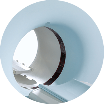
Modern imaging includes ultrasound, CT scanning, and MRI scanning.
Modern imaging has illuminated our understanding of TOS and our ability to diagnose TOS exponentially. Modern imaging has illuminated our understanding of TOS and our ability to diagnose TOS exponentially. Modern imaging has illuminated our understanding of TOS and our ability to diagnose TOS exponentially. Modern imaging has illuminated our understanding of TOS and our ability to diagnose TOS exponentially.

Modern imaging has illuminated our understanding of TOS and our ability to diagnose TOS exponentially. Modern imaging has illuminated our understanding of TOS and our ability to diagnose TOS exponentially. Modern imaging has illuminated our understanding of TOS and our ability to diagnose TOS exponentially. Modern imaging has illuminated our understanding of TOS and our ability to diagnose TOS exponentially.
Imaging of TOS
Modern imaging includes ultrasound, CT scan, and MRI scans, all useful in the diagnosis of TOS
Ultrasound has a place in the diagnosis and treatment of thoracic outlet syndrome. Ultrasound can demonstrate changes in blood flow in arteries and veins of the thoracic outlet in real time. It can be performed in almost any position of the chest, neck and arms, and in almost any angle relative to the anatomy of the area of interest.
Ultrasound has a place in the diagnosis and treatment of thoracic outlet syndrome. Ultrasound can demonstrate changes in blood flow in arteries and veins of the thoracic outlet in real time. It can be performed in almost any position of the chest, neck and arms, and in almost any angle relative to the anatomy of the area of interest.
Ultrasound has a place in the diagnosis and treatment of thoracic outlet syndrome. Ultrasound can demonstrate changes in blood flow in arteries and veins of the thoracic outlet in real time. It can be performed in almost any position of the chest, neck and arms, and in almost any angle relative to the anatomy of the area of interest.
Ultrasound has a place in the diagnosis and treatment of thoracic outlet syndrome. Ultrasound can demonstrate changes in blood flow in arteries and veins of the thoracic outlet in real time. It can be performed in almost any position of the chest, neck and arms, and in almost any angle relative to the anatomy of the area of interest.
Ultrasound has a place in the diagnosis and treatment of thoracic outlet syndrome. Ultrasound can demonstrate changes in blood flow in arteries and veins of the thoracic outlet in real time. It can be performed in almost any position of the chest, neck and arms, and in almost any angle relative to the anatomy of the area of interest.
Ultrasound has a place in the diagnosis and treatment of thoracic outlet syndrome. Ultrasound can demonstrate changes in blood flow in arteries and veins of the thoracic outlet in real time. It can be performed in almost any position of the chest, neck and arms, and in almost any angle relative to the anatomy of the area of interest.


