

Introduction
A single specialized examination can assess the structures involved in neurogenic, venous, and arterial TOS, including how they relate to surrounding anatomy and arm position.
Table of Contents
Table of Contents
- Three types of thoracic outlet syndrome share a crowded anatomical passage
- How the NeoVista MRI evaluates the thoracic outlet
- Neurogenic TOS and the search for a compression site
- Venous TOS and subclavian vein compression
- Arterial TOS and evaluation of the subclavian artery
- Why a combined examination can help
- Comparing the three types of TOS
- Frequently asked questions about NeoVista MRI and TOS
- Discussing NeoVista with your care team
- Sources
A single specialized examination can assess the structures involved in neurogenic, venous, and arterial TOS, including how they relate to surrounding anatomy and arm position.

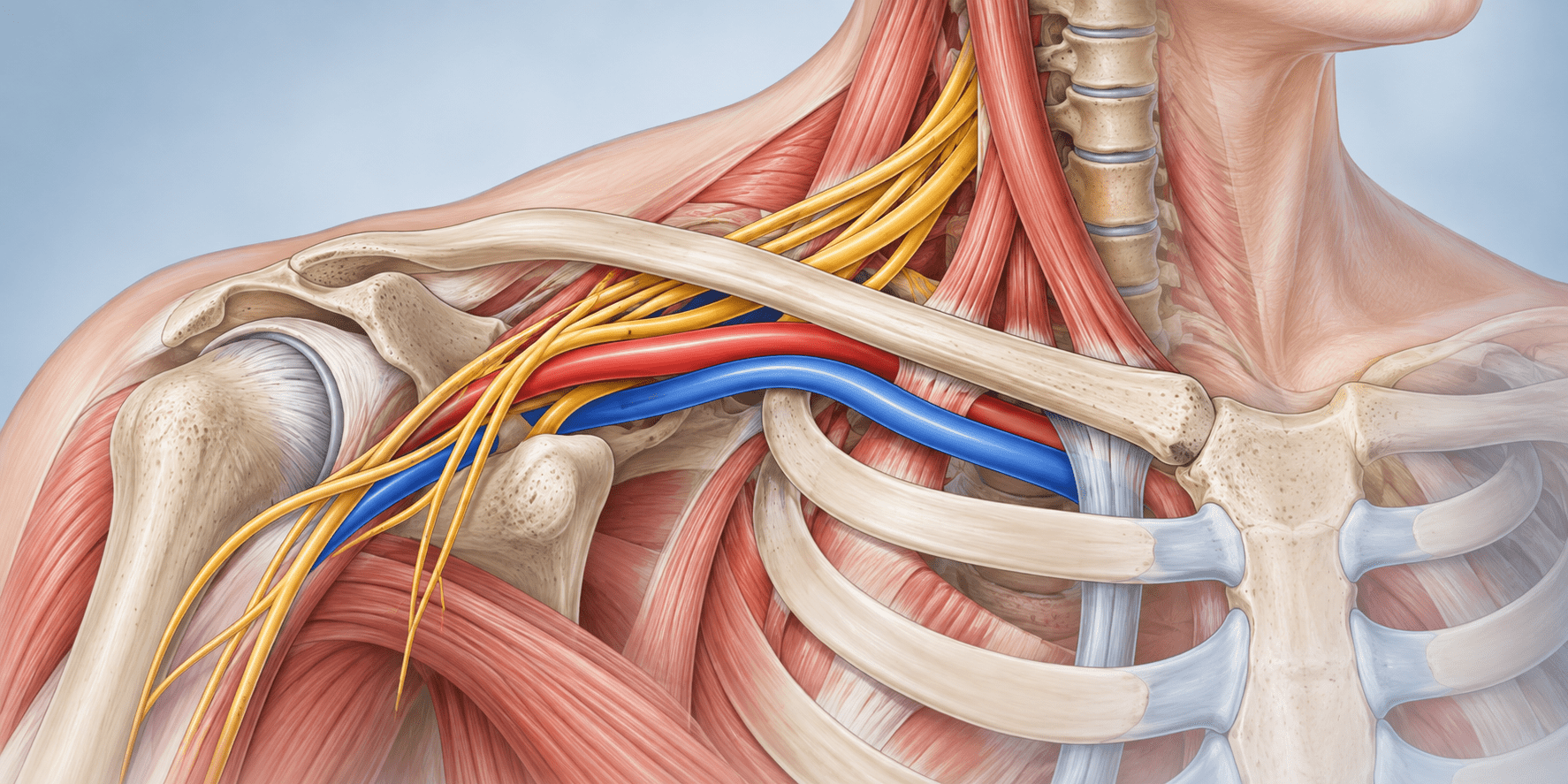
Illustration: nerves and blood vessels pass through the thoracic outlet beneath the collarbone.
By Vanguard Specialty Imaging | September 30, 2026
What is the NeoVista MRI for thoracic outlet syndrome? NeoVista® is a specialized MRI and MR angiography/venography examination developed to evaluate the thoracic outlet, where the brachial plexus and subclavian vessels pass between the neck and arm. Depending on the clinical question and protocol, it can assess both sides, show nerves, arteries, veins, bones, and soft tissues, and compare images acquired with the arms at rest and elevated. These findings help a clinician evaluate possible TOS and plan next steps; they are interpreted together with the patient’s history, examination, and other testing.
Three types of thoracic outlet syndrome share a crowded anatomical passage
Thoracic outlet syndrome (TOS) describes a group of conditions involving compression or injury to nerves or blood vessels as they pass from the neck and chest toward the arm. On each side, the brachial plexus, subclavian artery, and subclavian vein travel through a narrow space near the clavicle and first rib. The structure affected determines the main TOS subtype: neurogenic TOS affects the brachial plexus, venous TOS affects the subclavian vein, and arterial TOS affects the subclavian artery. Some patients may have overlapping findings.
Neurogenic TOS is the most common type. Pain, tingling, numbness, or weakness can overlap with cervical spine, shoulder, or peripheral nerve conditions, so evaluation often requires a careful clinical assessment and more than one kind of test. Venous TOS may cause arm swelling, heaviness, or bluish discoloration, especially when a subclavian vein clot is present. Arterial TOS is uncommon; when the artery has sustained injury, complications can include aneurysm, clot, or reduced blood flow to the hand. [1]
A routine neck or shoulder MRI may not answer the same questions as a dedicated thoracic outlet examination. TOS involves a specific anatomical region, several different structures, and in some cases changes associated with arm position. A targeted study is designed to show the relevant nerves, vessels, bones, and soft tissues together.
How the NeoVista MRI evaluates the thoracic outlet
NeoVista® is Vanguard Specialty Imaging’s dedicated MRI protocol for TOS. The protocol combines detailed anatomical sequences with MR angiography (MRA) and MR venography (MRV). Vanguard describes the examination as a roughly 45-minute study that uses no ionizing radiation and may use a tailored, diluted gadolinium contrast technique to image vessels on both sides from one injection. Contrast use and protocol details are determined by the clinical situation and the imaging team.
The central idea is to evaluate the neurovascular structures alongside the anatomy that may affect them. An angiographic image can show a vessel narrowing, for example, while anatomical images can help assess its relationship to the clavicle, first rib, scalene muscles, or other nearby structures. The examination also includes images in different arm positions when appropriate, because the thoracic outlet can change as the shoulder moves. Society for Vascular Surgery guidance likewise describes cross-sectional vascular imaging in neutral and elevated positions as one way to assess vascular entrapment and its relationship to nearby structures. [1]
According to Vanguard, the NeoVista examination can include evaluation of:
- The course and appearance of the brachial plexus on each side
- The subclavian arteries and their branches, including possible narrowing, aneurysm, or clot
- The subclavian veins and central chest veins, including possible narrowing, thrombosis, or collateral pathways
- The clavicles, first ribs, possible cervical ribs, and other bony variations
- Scalene muscles, pectoralis minor region, fibrous bands, and other soft tissues when visible
- Changes in anatomy and vessel caliber between arm positions, as specified by the protocol
- The cervical and upper thoracic spine when needed to assess alternative or coexisting findings
The complete protocol and the structures visible depend on the patient, scanner, and clinical question. Like any imaging method, MRI has limits. For example, the Society for Vascular Surgery notes that some ligamentous bands may be beyond the resolution of current imaging modalities. [1]
Neurogenic TOS and the search for a compression site
Neurogenic TOS can be difficult to evaluate because symptoms such as arm pain, tingling, numbness, and weakness can also occur with cervical radiculopathy, shoulder conditions, or peripheral nerve disorders. No single provocative maneuver, electrodiagnostic study, or imaging exam confirms every case. Clinicians use the patient’s history and examination together with testing selected for the individual. [1,2]
A dedicated MRI can add an anatomical view of the brachial plexus and surrounding structures. In particular, the study may assess three commonly discussed regions:
- Scalene triangle. The nerve roots and trunks pass between the anterior and middle scalene muscles. Imaging may show muscle or bony variants and the relationship of the plexus to nearby structures.
- Costoclavicular space. The plexus and vessels pass between the clavicle and first rib. Comparing positions may help show whether this space narrows with arm elevation.
- Subcoracoid or retropectoralis minor space. The neurovascular structures continue beneath the pectoralis minor region. Arm position and individual anatomy may affect this passage as well.
NeoVista also evaluates whether there are cervical ribs, elongated transverse processes, or other bony variations, and may include the cervical and upper thoracic spine to assess alternative or coexisting causes of symptoms. The scan is one part of the workup: clinicians interpret its findings in context, and a normal or nonspecific image does not by itself rule out every clinical form of neurogenic TOS.
Venous TOS and subclavian vein compression
Venous TOS involves the subclavian vein, commonly near the space between the clavicle and first rib. Repeated compression can injure the vein. In some patients, this is associated with effort thrombosis (Paget-Schroetter syndrome), which may cause sudden arm swelling, heaviness, or discoloration. Other patients have positional narrowing without a clot. [1,3]
Ultrasound is often used to evaluate suspected upper-extremity deep vein thrombosis. Additional vascular imaging may help answer questions about the location and extent of narrowing, surrounding anatomy, collateral veins, and possible changes with arm position. NeoVista’s MRV and anatomical sequences are intended to assess the vein together with nearby structures, with both sides evaluated as specified by the protocol.
Seeing the anatomy on both sides can provide useful context when symptoms are mostly on one side. However, positional vessel compression may also occur in people without symptomatic TOS. Imaging results therefore need to be interpreted with the patient’s symptoms and clinical evaluation rather than treated as a diagnosis on their own. [1,3]
Arterial TOS and evaluation of the subclavian artery
Arterial TOS is the least common subtype and is associated with compression or injury of the subclavian artery. A cervical rib or an unusual first rib can contribute in some cases. When arterial wall injury occurs, potential complications include post-stenotic dilation or aneurysm, mural thrombus, embolization, or reduced blood flow to the hand. These findings require timely assessment by a qualified clinician. [1,4]
NeoVista’s MRA and anatomical images can help show the subclavian arteries and their relationship to surrounding bone and soft tissue. The examination may assess the vessel at rest and with arm elevation, as well as its branches and signs of aneurysm, narrowing, or clot. The specific imaging approach and next steps depend on the patient’s presentation; urgent symptoms such as a suddenly cold, pale, painful, or weak hand require immediate medical attention.
Why a combined examination can help
TOS can involve more than one structure, and its symptoms can overlap with other conditions. A combined examination is intended to bring several kinds of information into one session: anatomy, nerve appearance, arterial and venous findings, and selected positional changes. This can give the ordering clinician and specialist a broader view than a test focused on only one structure or one position.
Vanguard reports that every NeoVista study is supervised and interpreted by Dr. Scott Werden, who developed the protocol, and that its reference library contains more than 3,000 TOS studies. Vanguard also states that it holds three U.S. patents relating to the imaging approach and describes its experience with surgical observation and clinical follow-up. These are Vanguard’s program details; the value of any individual study depends on image quality, interpretation, and how the result fits the clinical picture.
For referring clinicians, a dedicated study may help characterize anatomy, identify vascular complications, consider alternative diagnoses, and provide information for treatment planning. For patients, it may add useful information when symptoms persist or a workup has not yet explained them. It does not replace an examination by a clinician familiar with TOS, and it cannot promise a specific diagnosis or treatment outcome.
Comparing the three types of TOS
| Type | Main structure | Possible symptoms or findings | What imaging may assess |
|---|---|---|---|
| Neurogenic | Brachial plexus | Pain, tingling, numbness, or weakness | Plexus course and surrounding anatomy; possible compression or positional change |
| Venous | Subclavian vein | Arm swelling, heaviness, discoloration; possible clot | Vein narrowing, thrombosis, collateral veins, and nearby structures |
| Arterial | Subclavian artery | Hand coolness or discoloration; possible aneurysm or embolic complication | Artery narrowing, aneurysm, clot, branches, and bony relationship |
These categories describe the main structure involved. Symptoms and findings vary, and some patients may have overlapping conditions. The table is educational and is not a diagnostic tool.
Frequently asked questions about NeoVista MRI and TOS
Can an MRI diagnose thoracic outlet syndrome?
MRI can show anatomy and some abnormalities associated with TOS, but no single scan diagnoses every case. A clinician considers symptoms, examination findings, imaging, and other tests. This is especially important for neurogenic TOS, which can overlap with other nerve and musculoskeletal conditions.
What does a thoracic outlet MRI look for?
A dedicated examination may evaluate the brachial plexus, subclavian artery and vein, clavicle, first rib, nearby muscles and soft tissues, and changes associated with arm position. Protocol details vary by the clinical question and imaging center.
How is NeoVista different from a routine MRI?
NeoVista is a specialized TOS protocol that combines anatomical MRI with MR angiography and venography. Vanguard describes bilateral imaging and selected arm positions as part of its approach, so the study is designed to assess more than one structure in the thoracic outlet.
Does the NeoVista MRI use radiation?
MRI does not use ionizing radiation. The NeoVista protocol may use gadolinium-based contrast for vascular imaging. The ordering clinician and imaging team determine whether contrast is appropriate for the individual patient.
Does the examination use contrast?
Vanguard describes using a tailored diluted gadolinium technique for vascular images. Whether contrast is used and how it is administered depends on the clinical protocol and the patient’s circumstances; patients should discuss allergies, kidney disease, pregnancy, and prior contrast reactions with their care team.
Can NeoVista evaluate both sides of the body?
Vanguard’s protocol is designed to assess both thoracic outlets during one examination. The specific sequences and image quality depend on the scanner, protocol, and patient factors.
How long does a NeoVista MRI take?
Vanguard describes the examination as taking about 45 minutes. Actual appointment and scan times can vary by location and by the imaging needed.
Who should I talk to if I think I have TOS?
Start with a qualified clinician who can review your symptoms and examine you. Depending on the findings, they may refer you to a vascular, neurology, orthopedic, or other specialist and select appropriate testing. If you have sudden arm swelling or a cold, pale, painful, or weak hand, seek urgent medical care.
Discussing NeoVista with your care team
If you or your clinician are considering dedicated thoracic outlet imaging, ask what structures the examination will evaluate, whether imaging at different arm positions is appropriate, whether contrast is planned, and how the results will be interpreted alongside your clinical evaluation. Vanguard Specialty Imaging can provide information about the NeoVista MRI, imaging locations, and physician consultations through TOSMRI.com.
Medical information notice
This article is for general educational purposes and does not provide a diagnosis or replace medical advice. Imaging and treatment decisions should be made with a qualified healthcare professional who knows the patient’s history and symptoms.
Related reading: Imaging Every Form of Thoracic Outlet Syndrome: How the NeoVista® Protocol Works.
Sources
- [1] Society for Vascular Surgery. Thoracic Outlet Syndrome.
- [2] American College of Radiology. ACR Appropriateness Criteria: Thoracic Outlet Syndrome.
- [3] Likes K, et al. Venous thoracic outlet syndrome. Cardiovascular Diagnosis and Therapy.
- [4] Jones MR, et al. Thoracic Outlet Syndrome: A Comprehensive Review of Pathophysiology, Diagnosis, and Treatment.
- NeoVista MRI for Thoracic Outlet Syndrome. Vanguard Specialty Imaging.
- What Is the Best Test for Thoracic Outlet Syndrome? Vanguard Specialty Imaging.


